Neurocardiogenic
Cardiac—Arrhythmia
Channelopathies
Complete heart block
Sick Sinus syndrome
Tachyarrhythmias—supraventricular and ventricular tachycardia
Cardiac—Structural
Cardiomyopathy—hypertrophic/dilated
Coronary artery anomalies
Tumor
Left ventricular outflow obstruction
Primary pulmonary arterial hypertension
Eisenmenger syndrome
Mitral valve prolapse
Medication
Recreational (illegal)
Antiarrhythmic
Diuretics
Vasodilators
Producing QT prolongation
Neurologic
Seizure
Vertigo
Migraine
Tumor
Psychiatric
Conversion reaction
Panic attack
Hysteria
Hyperventilation
Metabolic
Hypoxia
Hypoglycemia
Diagnostic Evaluation
Given the many possible causes of syncope, the diagnostic evaluation can be quite involved and expensive and the specific etiology may never be determined. Therefore, a carefully planned approach rather than a “shotgun” diagnostic strategy is important. The patient history, family history, physical examination, and an electrocardiogram are fundamental and direct the remainder of the evaluation. Table 17.2 details the components of a comprehensive syncope evaluation. The patient history is the cornerstone on which the syncope evaluation is constructed and the diagnosis depends; it is often, along with the physical examination and an ECG, all that is necessary. Important historical details from the patient include: the age of the patient (syncope is rare before 10 years of age except for breath-holding spells in the toddler), time of day of the event (early morning is typical), the state of hydration and nutrition at the time of the event (when last had fluid or food intake), the environmental conditions (i.e., ambient temperature), the patient’s activity or body position immediately prior to the syncopal episode, the frequency and duration of the episodes, and any aura, prodrome, or specific symptoms and signs prior to the episode. Witnesses, if available, should provide details regarding the patient’s condition prior to the syncope, duration of loss of consciousness, any injuries or seizure-like movements, heart rate during episode (rarely available), and duration and nature of recovery (often patients are sleepy after neurocardiogenic syncope (NCS)). Medications (prescriptions and/or over-the-counter) used by the patient are critical historical points particularly regarding proarrhythmic agents such as QT prolonging medications. Information regarding prior diagnostic reports and/or consultations can prevent duplicate testing.
Table 17.2
Syncope evaluation
Patient historya |
Age at onset |
Time of day |
Frequency (increasing or decreasing over time) |
Prodrome (see text: patient may be amnestic) |
Situation (activity, place, body position) |
State of hydration |
Patient and witness description of event |
Duration of loss of consciousness |
Symptoms upon recovery (sleepy) |
Medications (prescription, OTC) |
Concomitant disease |
Prior evaluation and results |
Family history a |
Sudden death at young age |
SIDS |
Syncope |
Seizures |
Accidental death (e.g., drowning, automobile accidents) |
Pacemaker/defibrillator |
Congenital deafness |
Cardiomyopathy |
Patient exama |
General condition (habitus, phenotype, hydration, nutritional state, thyroid) |
Cardiac |
Blood pressure—sitting, lying, standing (for orthostatic response) |
Pulse—strength, rate, UE/LE difference |
Heart murmurs suggesting anatomic disease |
Musculoskeletal |
Inherited connective tissue disorder phenotype |
Neurologic |
ECGa |
Rate and rhythm |
AV conduction (heart block) |
Intraventricular conduction (Brugada syndrome, arrhythmogenic right ventricular dysplasia) |
QTc interval (male ≤ 0.45 s; female ≤ 0.46 s) |
T-wave morphology |
Exercise testingb |
Abnormal rhythm or blood pressure response. |
Especially rule out LQTS, exercise-induced syncope |
Noninvasive imagingb |
Echo Doppler |
Anatomic/structural assessment |
Tumors |
Pulmonary artery pressure estimate |
Outflow track gradients |
Abnormal origin left coronary artery |
Coronary aneurysms |
MRI |
Arrhythmogenic right ventricular dysplasia |
Coronary arteries |
Tumors |
Ambulatory monitoring |
Transtelephonic ECG |
Holterb |
Implantable recorder |
Head-up tilt testing (HUT)b |
Catheterizationb |
Hemodynamic |
Angiographic |
Electrophysiologic study |
Biopsy |
Right ventricular endomyocardial biopsy |
Family history is vital in the evaluation of syncope. It is not uncommon to find a history of multiple family members who experienced syncope during adolescence. Many of the older family members may also report a history of low blood pressure, and many families limit salt due to a hypertensive family member who is on a salt-restricted diet. However, if the family history is positive for recurrent syncope, it is also important to consider other familial disorders by specific questioning about the presence of hypertrophic or dilated cardiomyopathy, long QT syndrome (and other ion channelopathies), primary pulmonary hypertension, or arrhythmogenic right ventricular dysplasia. Families should be queried regarding sudden unexplained death in children or young adults (i.e., drownings, sudden cardiac death, sudden infant death syndrome, and car accidents), seizures, or familial congenital deafness (Table 17.2). Noting the person or source providing the family history and an estimate of its reliability can be of future use; it may be helpful to request further details from additional family members, particularly if a genetic disorder is suspected. A genetic counselor can be invaluable in helping to sort out the family history.
During the patient examination, orthostatic vital signs (magnitude of decrease in blood pressure relative to change from supine to erect position and heart rate changes) should be obtained. Many patients will manifest a mild (up to a 30 mmHg drop in blood pressure) but asymptomatic orthostatic change with upright positioning. An ECG should be obtained on every patient who experiences syncope, particularly if it is recurrent, occurs with exercise, and is not associated with the characteristic symptoms of NCS. The ECG should be evaluated for heart rate, corrected QT interval (gender-related), T-wave morphologic changes (persistent so-called juvenile T waves in the older adolescent), T-wave alternans, or any ventricular arrhythmia. The ECG should also be evaluated for ventricular preexcitation syndromes, atrioventricular (AV) conduction, or features of Brugada syndrome (see Chaps. 4, 13–15, 18).
For the infrequent patient whose evaluation lacks internal consistency, other studies may be advisable, such as echocardiography to examine for cardiomyopathy, myocarditis, anomalous coronary arteries, pulmonary arterial hypertension, or arrhythmogenic right ventricular dysplasia. A rare patient may warrant cardiac catheterization, including hemodynamic, angiographic, and electrophysiologic evaluation, along with right ventricular endomyocardial biopsy, to exclude potential structural, functional, and arrhythmic abnormalities, particularly before clearance to resume activities.
NCS: Simple Fainting, Vasovagal Syncope
Pathophysiology
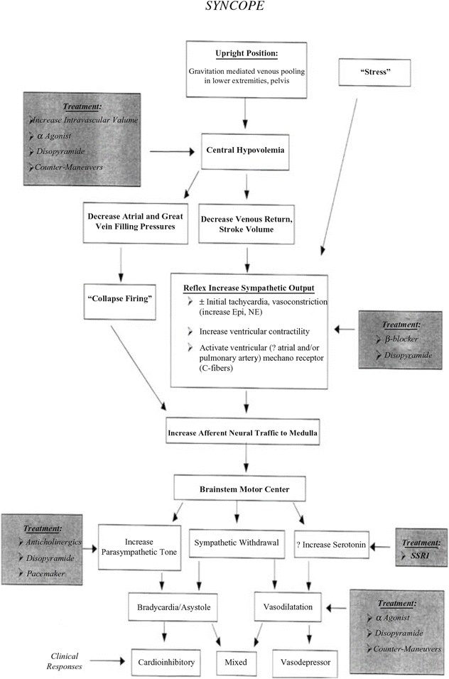
The pathophysiologic mechanisms underlying NCS are complex, likely heterogeneous, and not completely understood. However, one major hypothesis invokes a cardiac-central nervous system reflex (Fig. 17.1). The most common initiating event is prolonged (or the abrupt assumption of) upright position (sitting or standing), which subjects the patient to gravitationally mediated venous pooling in the lower extremities and pelvis. This causes an abrupt central hypovolemia (compared to the immediate preexisting state), leading to a decrease in venous return and stroke volume. In addition, an emotional or physical stress (e.g., pain or fright) or a reflex mechanism related to hair grooming, glutition (swallowing), or micturation may initiate this sequence by stimulating a reflex increase in sympathetic output manifested as tachycardia and vasoconstriction along with an increase in ventricular contractility. Activation of C-fiber mechanoreceptors increases afferent neural traffic to the central nervous system (medulla), stimulating the brain-stem motor center and causing several and often combined possible responses, such as the following: (1) an increase in parasympathetic activity, causing profound bradycardia or asystole; (2) a sympathetic withdrawal resulting in peripheral vasodilatation (venous and arterial), a decrease in systemic blood pressure, and decrease in heart rate; and (3) an increase in serotonin concentration, also resulting in peripheral vasodilatation and marked decrease in the systemic blood pressure. This sequence of events is one hypothesis, but it does not account for all the observed complex and integrated interaction between the neurohumoral traffic and the cardiovascular responses, additionally confounded by age and comorbidity.


Fig. 17.1
Algorithm of one possible mechanism for neurocardiac syncope [Reprinted from Strieper MJ. Distinguishing benign syncope from life threatening cardiac causes of syncope. Semin Pediatr Neurol 2005;12:32–38. With permission from Elsevier]
As a result of the loss of consciousness, postural tone and the upright state, the patient falls to a supine position restoring venous return and the central circulating blood volume (i.e., heart and lungs) followed by rapid normalization of blood pressure and heart rate. The loss of consciousness is usually short (generally ≤1–2 min). Excretory incontinence is uncommon. Seizures rarely occur as a result of the sudden prolonged decrease in cerebral perfusion. During recovery, return to sentience is rapid but post-event fatigue is common.
Clinical Presentation: NCS–NMS and Other Considerations
A prodrome lasting from several seconds to 1–2 min and consisting of nausea, epigastric discomfort, a clammy and cold sweat, pallor, dizziness, lightheadedness, tunnel vision, headache, and weakness is highly characteristic and strongly suggests simple fainting, vasovagal or neurocardiogenic syncope. If the prodrome is of sufficient duration, patients may learn to recognize their symptoms and lie down to relieve the symptoms and prevent syncope. Some patients with profound bradycardia or asystole may have little or no prodrome, causing a sudden loss of consciousness that may result in injury. Absence of a significant prodrome also raises the possibility of structural, functional, or arrhythmic causes for syncope. On the other hand, palpitations, chest discomfort, and a sudden loss of consciousness as well as a prompt recovery are more compatible with an isolated cardiac event. Other symptoms such as atypical precordial chest pain or tightness in the chest, breathlessness, acrocyanosis, tingling in the hands or feet, and a sense of alarm or anxiety are compatible with hyperventilation and a “panic attack.”
If “seizures” (tonic-clonic movements) occur as a result of cerebral hypoperfusion and anoxia, the patient can be confused as having a primary neurologic abnormality. Formal neurologic consultation and neurologic testing should be considered if seizures are part of the presentation. Interestingly, longstanding complaints of intermittent abdominal pain and nausea, most likely due to a profound increase in vagal tone, constitute an unusual and rarely suspected presentation of NCS. This symptom complex, as a manifestation of NCS, may be associated with a positive head-up tilt (HUT) evaluation, and may respond favorably to NCS treatment. In addition, there may be a link between NCS and chronic fatigue syndrome, which also has findings of hypotension, headaches, and postexertional fatigue. However, it is unlikely that an otherwise healthy preadolescent child or adolescent would exhibit the chronic fatigue syndrome.
Patients who present with syncope during exercise are also a challenge. Gradual loss of consciousness, associated with a pre-syncopal prodrome and occurring as the exercise is reaching its completion and the exerciser is in an exhausted state at the finish after maximal effort suggests the possibility of exercise-induced NCS, provoked by preexisting unrecognized dehydration, exercise-induced catecholamine-enhanced ventricular contractility, a sudden decrease at the end of exercise in the peripheral blood flow from the vasodilated skeletal muscle vasculature and cerebral vasoconstriction (induced by the respiratory alkalosis of the exercise-related hyperventilation) producing a decrease in cerebral blood flow at that critical moment and syncope. On the other hand, sudden syncope during exercise and without prodrome raises the suspicion for a more serious underlying cardiac structural or functional cause, including arrhythmias, and warrants further investigation.
< div class='tao-gold-member'>
Only gold members can continue reading. Log In or Register to continue
Stay updated, free articles. Join our Telegram channel

Full access? Get Clinical Tree


