Fig. 15.1
Sinus rhythm with a junctional premature beat (J), which results in AV node refractoriness and block of the next sinus beat. *Subsequent non-conducted atrial impulse may be due to concealed junctional extrasystole, again making the AV node refractory without activating atrium or ventricle. Proof of this phenomenon requires intracardiac recordings
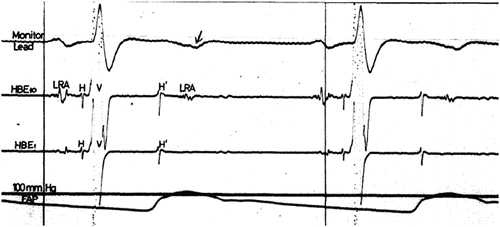
Fig. 15.2
Spontaneous His bundle extrasystoles (H′) arising at an H–H′ interval of 257 ms producing bigeminal rhythm. H′ fails to conduct to the ventricle but conducts retrograde to the atrium resulting in reversal of atrial activation from low to high atrium, associated with an inverted P wave (arrow). The bigeminal His bundle extrasystoles cause a marked slowing of ventricular rate. [Reprinted from Nasrallah AT, Gillette PC, Mullins CE, et al. Concealed his bundle extrasystoles in congenital heart disease. American Journal of Cardiology. 1975;35(2):288–292. With permission from Elsevier.]
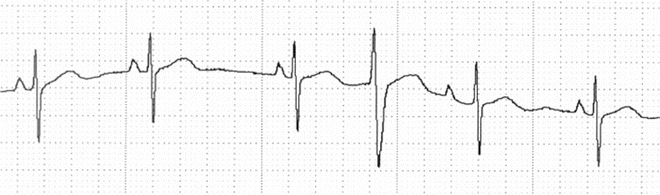
Fig. 15.3
Sinus rhythm with interpolated PVC. Concealed retrograde conduction into the AV node results in relative refractoriness and first-degree block (longer PR interval) of subsequent sinus impulse
Similar to the sinus node, the AV node is innervated by the autonomic nervous system, which consists of a complex interaction of the sympathetic and parasympathetic nervous systems.
Cardiac chronotropic and inotropy are influenced by differing sympathetic and parasympathetic effects. Sympathetic nerves descend from the brain to the stellate and paravertebral ganglia where they synapse with postganglionic neurons. The sympathetic nervous system increases heart rate and contractility by binding norepinephrine to adrenergic receptors, initiating the adenylate cyclase-cAMP cascade. Parasympathetic nerves originate in the brain stem and project from the vagus nerve to postganglionic fibers within cardiac ganglia. The parasympathetic nervous system slows heart rate and decreases contractility by binding acetylcholine to cardiac muscarinic receptors or neural nicotinic receptors. Postganglionic sympathetic nerves extend from the paravertebral ganglia to the heart where they meet with the parasympathetic nerves to form the cardiac neuronal plexus at the base of the heart.
The sympathetic innervation of the conduction system dominates in infancy but shifts to a balance of sympathetic and parasympathetic innervation in childhood and is complete in adulthood. Autonomic innervation is less prominent in the His–Purkinje system and has less influence on conduction in these areas. Bradycardia and PR interval prolongation usually reflects increased vagal tone on the sinus and AV nodes. PR interval prolongation in the presence of normal sinus rates is suggestive of AV nodal dysfunction. Functional first- and second-degree AV block can also present during rapid atrial pacing (Fig. 15.4), where, unlike sinus tachycardia, there is less sympathetic enhancement of AV nodal conduction.
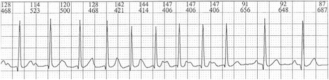
Fig. 15.4
Atrial pacing (stimulus artifact not present) producing a tachycardia that transitions to sinus rhythm. There is first-degree AV block during the tachycardia that resolves with the return of sinus rhythm
ECG Characteristics
Multiple channel ECG recordings are often indispensable for detection of P wave morphology and PR intervals. Recording of atrial activity from esophageal or temporary epicardial pacing leads after surgery (Fig. 15.5) are also very useful when P waves are indistinct. Long-term recordings from Holter monitoring often reveal patterns not apparent in the 12 lead electrocardiogram.
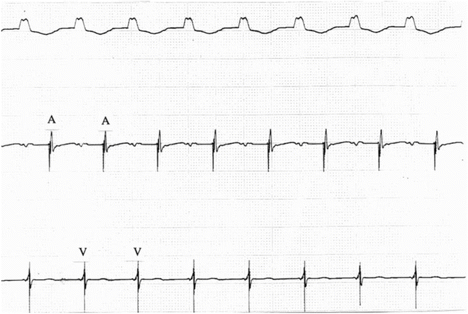
Fig. 15.5
Surface ECG (upper signal), atrial wire recording (middle signal), and ventricular wire recording in a patient with first-degree AV block following surgical repair of an atrioventricular septal defect. Atrial activity was not apparent from the surface ECG. Atrial wire recordings confirm 1:1 AV relationship with prolonged AV conduction time
First-Degree AV Block
First-degree AV block (Fig. 15.6) is defined as a PR interval above the normal range for age, but with persistent 1:1 AV conduction. The normal PR interval also decreases with increasing heart rate. Age-appropriate PR intervals are summarized in Table 15.1. The PR interval can be very long (Fig. 15.7) but usually, in the absence of heart disease, does not progress. The delay is located in the AV node mediated through excessive parasympathetic tone. Exercise, both recreational and during stress testing, induces parasympathetic withdrawal and sympathetic enhancement resulting in normalization of AV conduction and the PR interval (Fig. 15.7).
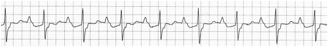
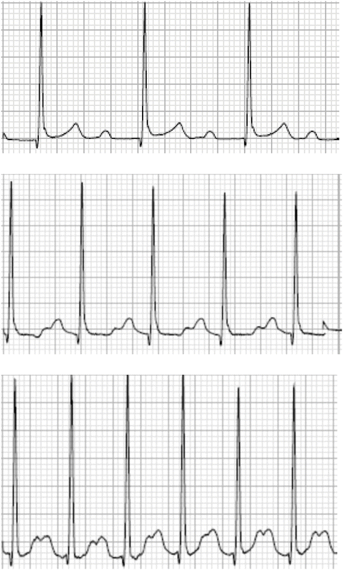
Fig. 15.6
Sinus rhythm with first-degree AV block
Table 15.1
Maximum normal PR interval
Age | PR (s) |
|---|---|
0–3 days | 0.16 |
4–30 days | 0.14 |
1–3 months | 0.13 |
4–6 months | 0.15 |
7–12 months | 0.16 |
1–5 years | 0.16 |
6–12 years | 0.17 |
>12 years | 0.20 |
Fig. 15.7
16-year-old girl with first-degree AV block at baseline. With exercise, the PR interval shortens and there is 1:1 AV conduction
Second-Degree AV Block
Several distinct patterns of second-degree AV block can be recognized. Consistent periodicity (i.e., dropping every third, fourth, or fifth beat) is frequently present. The ratio of P-to-R waves provides a description of the pattern (i.e., 3:2, 4:3). This pattern of “grouped beats” should always suggest second-degree AV block. Regular non-conducted atrial extrasystoles may also present with this pattern and are distinguished by the irregular PP interval, as well as the different P wave morphologies (Fig. 15.8).
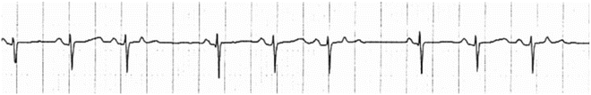
Fig. 15.8
Sinus rhythm with frequent non-conducted atrial extrasystoles in a quadrigeminal pattern. The grouped beats mimic second-degree AV block, but the variation in P wave timing and morphology are distinguishing features
Mobitz I (Wenckebach)
With typical Mobitz I, or Wenckebach (Figs. 15.9 and 15.10), there is gradual prolongation of the PR interval prior to a non-conducted beat. The greatest increase in PR interval is between the first and second conducted beats of a series. The lesser increment increase of the PR interval on subsequent beats leads to a shortening of the RR interval. Following the non-conducted beat, the normal PR interval is restored resulting in an RR interval that is less than twice the sinus rate.
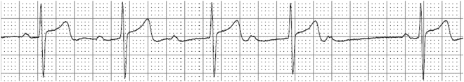
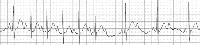
Fig. 15.9
Sinus rhythm with first-degree and Mobitz I second-degree AV block with a 5:4 conduction ratio
Fig. 15.10
Sinus rhythm with Mobitz I second-degree AV block that transitions to 2:1 block
Atypical Wenckebach, which may be more common than the typical form, refers to other patterns of PR prolongation in association with the appearance of a dropped beat (no conduction to the ventricles)—AV block. This pattern may occur in the presence of sinus arrhythmia and with longer runs of conducted beats between block cycles.
Mobitz II
The PR interval does not vary prior to non-conducted beats with Mobitz II second-degree AV block, and, in the absence of a supraventricular arrhythmia, the RR interval is constant (Fig. 15.11). Since there is no change in PR or RR interval during the conducted beats, the RR interval following a non-conducted beat should be twice that of a conducted RR interval.
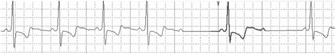
Fig. 15.11
Sinus rhythm with stable PR interval that transitions to 2:1 block. In the absence of prior PR interval prolongation, this most likely represents Mobitz II AV block
When every other beat is non-conducted in a 2:1 pattern, insufficient information is present to distinguish the pattern of Mobitz I from Mobitz II. Long-term recordings may reveal other ratios of conduction allowing discrimination of these two entities (Figs. 15.10 and 15.11). Long QT syndrome may present with 2:1 conduction as the ventricles, due to the mutated K+ ion channel (LQTS1) delaying ventricular repolarization, are refractory to successive sinus impulses; it is a poor prognostic sign.
Advanced
Advanced AV block is present when two or more impulses are not conducted in the absence of complete heart block (Fig. 15.12). Patients with advanced AV block may progress to complete heart block (Chap. 16).
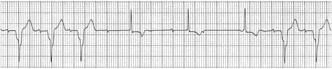
Fig. 15.12
Sinus rhythm in a patient with nodoventricular pathway. Advanced AV block associated with narrow complex escape rhythm
Electrophysiologic Features
Electrophysiologic studies have provided invaluable insights into AV node physiology. Currently, clinical history and noninvasive diagnostic studies primarily guide diagnosis and management of first- and second-degree AV block. Electrophysiologic study is not routinely performed solely for the assessment of first- and second-degree AV block because surface electrocardiograms provide adequate information for management. Invasive electrophysiologic studies of AV node function can be performed as an adjunct to hemodynamic study when deemed useful.
< div class='tao-gold-member'>
Only gold members can continue reading. Log In or Register to continue
Stay updated, free articles. Join our Telegram channel

Full access? Get Clinical Tree


