Surgical Reconstruction of the Supra-Aortic Trunks and Vertebral Arteries

Chapter 19 Surgical Reconstruction of the Supra-Aortic Trunks and Vertebral Arteries


The supra-aortic trunks (SATs) are the branches of the aortic arch that ascend through the mediastinum and terminate short of the carotid bifurcation and the origin of the vertebral arteries. These trunks carry the entire blood supply to the head and upper extremities. The vertebrobasilar system is composed of the two vertebral arteries, the basilar artery, and their branches to the brainstem, spinal cord, cerebellum, and occipital lobes.


The three most common variants in the anatomy of the SATs have implications in terms of the technique chosen for their reconstruction. These variants are a shared ostium (16%) or a common origin (8%) for the innominate and left common carotid arteries, a left vertebral artery with a separate origin from the aortic arch (6%), and a right retroesophageal subclavian artery (0.5%). A separate origin of the left vertebral artery is associated with an abnormally high (C4 or C5) entry of this artery into the transverse foramina of the cervical spine. A retroesophageal right subclavian artery is associated with a thoracic duct that empties on the right jugulosubclavian confluent, a nonrecurrent right inferior laryngeal nerve, and, in approximately half these patients, a common carotid trunk giving origin to both common carotid arteries.


Through the mechanisms of low flow or atheroembolization, occlusive disease of the SATs can cause symptoms in any of the territories supplied: the hemispheric (carotid) territory, the posterior (vertebrobasilar) territory, and, in the case of proximal subclavian disease, the upper extremity. Vertebral artery occlusive disease may restrict inflow into the basilar artery, resulting in vertebrobasilar ischemia. This is more likely if compensatory flow from the carotid system is not available because of an internal carotid occlusion or a minute or absent posterior communicating arteries.


The SATs are involved by atherosclerosis in the fifth or sixth decade of life. This results in the development of plaques that can obstruct flow or embolize (atheroembolism). Aneurysmal atherosclerotic disease of the SATs is rare. In some locations, the SATs are a common site for Takayasu arteritis, usually in younger individuals. Traumatic and mycotic aneurysms of the SATs are uncommon but life-threatening conditions.


The incidence of atherosclerotic disease is lower in the SATs than in the internal carotid and vertebral arteries. Nevertheless, the extensive study of extracranial arterial disease reported in 1968 by Hass and colleagues1 showed that one third of patients undergoing arteriography had a severe lesion involving one or more of the SATs. The morphology of atherosclerotic lesions of the SATs is not defined as well as that of the plaques found in the internal carotid artery, partly because for several years the SATs were not routinely visualized during arteriography of the cerebral vessels. In addition, the SATs are difficult to image with ultrasound techniques, which provides valuable morphologic information in the carotid bifurcation. Because there is limited knowledge of the natural history of these lesions, surgical indications are based partly on inferences. In addition, SAT lesions are often found in individuals who already have concomitant disease of the carotid or vertebral arteries—a situation that confuses the identification of the offending lesion. Stenosing lesions of the SATs usually appear at their origin from the aortic arch and often involve more than one artery. Plaques located in the ostia of the SATs are usually continuous with atheroma that extends over the dome of the aortic arch.


Outlining SAT lesions by arteriography requires an arch injection, preferably in two oblique projections (right and left posterior oblique). If circumstances permit, the arteriographic study should also include selective injections of both common carotid and subclavian arteries (four-vessel arteriogram). The high incidence of concomitant carotid and vertebral artery lesions makes it mandatory to outline the extracranial and intracranial cerebrovascular supply when evaluating a patient for cerebrovascular symptoms.


Vertebrobasilar ischemia can be caused by poor inflow through the carotid and vertebral arteries, reversal of blood flow in a vertebral artery caused by a proximal subclavian artery occlusion (subclavian steal), reversal of right carotid and vertebral artery flow from an innominate artery occlusion, and embolization from the proximal subclavian or vertebral arteries.


Symptoms of Occlusive Disease of the Supra-Aortic Trunks


Patients with occlusive disease of the SATs may show symptoms of carotid, vertebrobasilar, and upper extremity arterial ischemia. Although there is no pathologic evidence to support this view, it has traditionally been taught that in patients with disease of the SATs, cerebral symptoms are due to low flow rather than atheroembolization. This concept runs contrary to clinical evidence suggesting that the mechanisms of cerebral ischemia from disease of the SATs are similar to those from atheroma of the carotid bifurcation.


Obliteration of the SATs is suggested by absent pulses in the neck (subclavian, carotid) or arm (axillary, brachial) on one or both sides and by the recording of unequal or abnormally low pressures in the upper extremities. Waveforms recorded by Doppler tracings are dampened in arteries whose origins are stenosed or occluded. Bruits may be present. In patients with subclavian steal, a pulse lag may be felt between the radial arteries of the two arms or, more precisely, a pulse wave delay of greater than 30 msec can be measured by simultaneously recording both brachial artery waveforms.2 Claudication of the arm and digital artery embolization may be present in subclavian artery disease. A computed tomographic scan of the brain is an essential part of the workup for patients with disease of the SATs. The scan may reveal clinically silent cerebral infarctions.3


In symptomatic vertebral (or basilar) artery occlusive disease, the patient may have any combination of the following symptoms: dizziness, vertigo, diplopia, perioral numbness, blurred vision, tinnitus, ataxia, bilateral sensory deficits, and drop attacks. The mechanism that triggers the symptom (e.g., standing up, rotating the neck) must be sought when evaluating these patients. Patients with orthostatic hypotension have vertebrobasilar symptoms when they stand abruptly after sitting or lying down. Blood pressure measurements taken immediately after they stand up and experience symptoms shows a drop in systolic pressure greater than 20 mm Hg. This mechanism is particularly common in diabetic patients with sympathetic paralysis leading to loss of venomotor tone because a substantial amount of blood is pooled in their legs on standing.


The presence of vertebrobasilar ischemia related to turning of the neck suggests osteophytic compression on the vertebral arteries or inner ear disease. In general, in patients with labyrinthine disorders, symptoms appear with brief, head-shaking motions. Patients who develop symptoms by extrinsic compression of the vertebral arteries usually require a few seconds with the neck rotated maximally in a particular direction to develop symptoms. In addition to orthostatism and osteophytic compression, other conditions are capable of causing vertebrobasilar ischemia and must be ruled out. Dissection of a vertebral artery is accompanied by neck pain. In these patients, the symptoms of brain ischemia may be due to critical compromise of the true lumen of the vertebral artery by an intramural dissecting hematoma or thromboembolization from the distal reentry point of the dissection. A number of medical conditions also present with symptoms of vertebrobasilar ischemia; among the most common are inappropriate antihypertensive medication, cardiac arrhythmia, anemia, brain tumor, and subclavian steal.


Indications for Surgery


No morphologic database exists for atherosclerotic lesions of the SATs comparable to that available for internal carotid artery disease. It is known from arteriograms and postmortem studies that the SATs are less frequently involved by atherosclerotic disease than is the carotid bifurcation. In general, clinicians do not have the ability to use ultrasonography to study the composition of SAT plaques, and the specimens obtained at operation are few because the interventions to reconstruct the SATs are bypasses rather than endarterectomies. The few specimens available for pathologic study show degenerative features similar to those seen in carotid plaques: surface thrombus, ulceration, and intraplaque hemorrhage. One may reasonably infer that the same pathologic mechanisms operate in both carotid artery plaques and SAT lesions. Until more precise information becomes available, it seems sensible to use criteria similar to those applicable to carotid disease to determine treatment guidelines. These criteria, however, must be tempered by the fact that the risk of surgical reconstruction of the SATs is higher than that of carotid endarterectomy.


Indications for surgical repair of SAT lesions are (1) lesions encroaching on more than 70% of a SAT diameter, or plaques with ulceration or surface irregularities in patients with appropriate symptoms (ipsilateral carotid or vertebrobasilar); (2) the same lesions plus ipsilateral internal carotid disease for which an endarterectomy is indicated (the operation should correct both); (3) the same lesions plus a nonacute ipsilateral hemispheric infarction (overt or silent); and (4) preocclusive lesions (>90% cross-sectional area loss) in asymptomatic patients who are good surgical risks and have more than 5 years of life expectancy. This last indication is arbitrary albeit reasonable.


The primary indication for reconstructing a vertebral artery is to treat vertebrobasilar ischemia. Severe occlusive disease of the vertebral artery may be found in individuals who have no symptoms of vertebrobasilar ischemia and do not require surgical treatment. Conversely, many systemic causes of vertebrobasilar ischemia are not related to vertebral artery disease. Therefore the decision to reconstruct a vertebral artery must be based on a strong anatomic and clinical presumption that the symptom (vertebrobasilar ischemia) is secondary to the anatomic lesion (occlusive disease of the vertebral arteries).


Vertebrobasilar ischemia may be due to stenosis or occlusion of the vertebral or basilar arteries, restricting flow in the territory supplied by these arteries. This is the so-called low-flow (or hemodynamic) mechanism. These patients often have repetitive transient ischemic attacks triggered by positional or postural mechanisms. Although their risk for stroke is lower than that in patients with carotid disease, they may suffer serious traumatic injuries because of loss of balance (such as syncopal attack while driving). In patients with low-flow symptoms of ischemia in the vertebrobasilar territory, the surgical indication rests on the assumption that the basilar artery is not receiving adequate inflow from the vertebral arteries. Ischemia of the vertebrobasilar territory may also be due to microembolization (atheroemboli). Contrary to prevalent views in the neurologic literature, approximately one third of vertebrobasilar ischemic episodes are caused by atheroembolization from plaques or mural lesions of the vertebral arteries.4 Patients with embolic symptoms are at high risk for infarctions of the brainstem, cerebellum, and occipital lobes. The mechanism here is microembolization from the irregular surface or from the core of a plaque in the proximal subclavian or vertebral arteries or from a lesion in the wall of the vertebral artery secondary to repetitive trauma from an osteophyte or from intramural dissection.


In patients with low-flow symptoms, and because two vertebral arteries usually supply the basilar artery, the presence of a normal vertebral artery contraindicates an operation on the opposite artery, regardless of its anatomic condition. A vertebral artery of normal caliber emptying into a basilar artery is enough to supply the basilar territory. This means that for a lesion in the vertebral arteries to be considered significant, it must be severe (>75% stenosis) and the opposite vertebral artery must be equally diseased, hypoplastic, or absent.


My approach to a patient with low-flow vertebrobasilar ischemia is first to determine whether any other clinical condition (e.g., orthostatism, arrhythmia) capable of producing these symptoms is present. If so, it should be corrected. If symptoms persist after treatment, an arteriogram is indicated. If the arteriogram shows a lesion that fulfills the anatomic criteria listed previously and the operation appears to be technically feasible, a reconstruction of the vertebral artery is indicated.


In patients with vertebrobasilar ischemic symptoms secondary to embolization, the indication for surgery rests on demonstration of the emboligenic lesion, regardless of the condition of the opposite vertebral artery. The criteria of bilateralism and degree of severity that apply to low-flow lesions are irrelevant when considering treatment for atheroembolic disease.


Reconstruction of the Supra-Aortic Trunks


The main decision involved in reconstruction of the SATs is whether to do the repair through the chest or through the neck. Cervical repairs are traditionally done by means of a bypass from a suitable donor vessel to the diseased one. Most of these bypasses run transversely either between vessels on the same side of the neck (carotid and subclavian) or across the neck (remote bypasses). Bypass procedures between the ipsilateral carotid and subclavian arteries are largely being superseded by transposition procedures that provide the advantage of a single arterial anastomosis without the need for a saphenous vein graft or a prosthetic tube. Transthoracic or axial repairs require a partial or total sternotomy for a direct approach to these vessels. The lesions are corrected with a bypass from the ascending aorta.


The choice between transthoracic (axial) and cervical (transverse) repairs can be made using the following general guidelines. Axial repairs are preferred in younger patients who have innominate artery lesions or multiple lesions (usually innominate and left common carotid). They are also the natural choice for patients in whom a simultaneous coronary bypass operation is indicated. In patients with atherosclerotic disease of the SATs and coronary arteries, repairing concomitant severe lesions of the first segment of the left subclavian artery is advisable even if this stenosis is asymptomatic. This repair permits a later myocardial revascularization using the left internal mammary artery.


Cervical repairs are preferred in older patients, in those who are at high risk of thoracotomy, and in those who have had previous transsternal procedures. Cervical repair is the choice for all single arterial lesions (other than those of the innominate artery).


Cervical Repairs


In the early 1970s, techniques for revascularization of the SATs consisted of a transverse bypass between a donor and a recipient (diseased) artery. The insertion of a bypass between the carotid and subclavian arteries, although described in 1957,5 did not become popular until the 1970s. These bypasses were between the midsegment of the carotid artery and the second (retroscalene) segment of the subclavian artery. In some cases, the carotid artery acted as the donor vessel, and the bypass corrected a blockage of the first portion of the subclavian artery. In others, the subclavian artery was the donor vessel to bypass a proximal common carotid artery lesion. In subclavian-carotid bypasses, when the anastomosis to the carotid artery is of the end-to-side type, there is always the possibility of a source of proximal embolization (from the diseased proximal common carotid artery) or of extension of the proximal thrombus across the end-to-side anastomosis. As a result, an end-to-end anastomosis (see later) into the common carotid artery is recommended.


Carotid-subclavian bypasses became the standard operation for the correction of subclavian steal syndrome in the 1970s. When use of the carotid as the donor vessel was not advisable (because it was the only patent carotid artery or because of disease in the proximal common carotid artery), correction of subclavian steal was achieved with bypasses between both subclavian arteries or both axillary arteries. These remote bypasses became known as extraanatomic operations. Although they offer the advantages of avoiding a thoracotomy and carrying a lower operative mortality, they have lower long-term patency rates than those achieved by axial or shorter reconstructions. These remote bypasses are constructed with the graft crossing the neck in front of the sternum, giving a poor cosmetic result and subjecting them to external compression. Most significantly, bypasses in this anterior, low-neck location may interfere with an eventual tracheostomy and certainly with a midsternotomy that might be required for coronary revascularization later on.


Many of the cervical bypasses done in the 1970s used saphenous vein as the preferred graft material. There was fear of embolization from the “neointima” of prosthetic tubes and doubts about their long-term patency. However, saphenous veins also presented specific problems. They were not always available, and gross mismatches in caliber often occurred between the vein and the recipient arteries. In addition, the length required for remote bypasses led to the possibility of axial rotation or compression or kinking of the vein graft with rotation of the neck. Because of these difficulties, surgeons explored the use of prosthetic substitutes for arterial bypass in the neck. The reported results indicate that polytetrafluoroethylene (PTFE) grafts are preferable to saphenous vein for bypasses in the neck; they provide a good-caliber match, and their patency rates are excellent,5 probably as a result of the high flow rates usually measured in these arteries.


Anatomic Indications


Common Carotid Artery Occlusion or Stenosis.


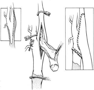
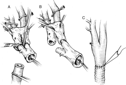
The common carotid artery can be revascularized by means of a subclavian-carotid bypass from the ipsilateral subclavian artery, with the distal anastomosis being performed end to end to avoid embolization from the diseased proximal common carotid artery. When the entire carotid system on one side is not visualized on the arteriogram, one must consider the possibility that the carotid bifurcation is patent, with retrograde flow from the external carotid artery perfusing the internal carotid artery antegradely. Delayed subtraction films may show this late opacification, but duplex imaging is the best way to show patency of the carotid bifurcation and this peculiar combination of retrograde (external carotid) and antegrade (internal carotid) flow. In these patients, atheromatous plaque is usually found at the origin of the internal carotid artery, necessitating endarterectomy before performing the distal anastomosis. The traditional means of constructing a distal anastomosis for a graft at the level of the carotid bifurcation is with an end-to-side junction (an onlay patch), with or without a concomitant endarterectomy of the carotid bulb (Figure 19-1). This end-to-side anastomosis is functionally transformed into an end-to-end junction by ligation of the common carotid artery below the anastomosis, in the soft segment created by endarterectomy of the distal portion of the common carotid artery. A preferable technique for the distal anastomosis is (Figure 19-2): to amputate of the distal common carotid artery, the carotid bulb is then everted for endarterectomy (type 2 eversion). The round cross section of the carotid bifurcation allows a simple end-to-end anastomosis to the bypass arising from the subclavian artery.


If the stenosing lesion of the common carotid artery is located at its origin and its distal two thirds are free of disease, transposing the midportion of the common carotid artery to the subclavian artery is a better solution than a subclavian-carotid bypass. It requires only one anastomosis and no prosthesis. At times, a thrombosed common carotid artery with a patent bifurcation can be thrombectomized after dividing it low in the neck and doing an eversion endarterectomy up to the bifurcation. The distal portion of the endarterectomy is terminated under direct vision through the standard arteriotomy used for a conventional carotid endarterectomy. After endarterectomy, the common carotid artery is reimplanted into the second portion of the subclavian artery. Subclavian-carotid bypass and transposition of the carotid into the subclavian are easier on the right side, where the subclavian artery is more accessible.


In cases in which the ipsilateral subclavian artery is not a suitable donor vessel, a common carotid artery lesion can be corrected by means of a carotid-carotid bypass. This operation is traditionally done by placing a bypass between both carotids in front of the airway. Instead, I prefer the shorter retropharyngeal route (see later in this chapter).


< div class='tao-gold-member'>

Stay updated, free articles. Join our Telegram channel

Jul 1, 2016 | Posted by in CARDIOLOGY | Comments Off on Surgical Reconstruction of the Supra-Aortic Trunks and Vertebral Arteries

Full access? Get Clinical Tree

Get Clinical Tree app for offline access