Surgery for Atrial Fibrillation
The Maze procedure was developed and modified by Dr. James Cox and has proved to be effective for treating atrial fibrillation associated with valvular and ischemic heart disease and isolated atrial fibrillation refractory to medical therapy. The Maze III cut and sew technique is the gold standard against which modifications should be measured because of its greater than 95% cure of atrial fibrillation. However, this procedure adds significantly to the aortic clamp time and incurs the risk of serious bleeding from the back of the heart. Several different energy sources have been used to ablate atrial tissue creating the same lesion pattern as the Maze III operation in less time and with less bleeding potential. We have used an irrigated radiofrequency probe, but the same ablation lines can be accomplished with cryoablation, microwave energy, focused ultrasonography, or laser. Patients with chronic atrial fibrillation undergoing mitral valve surgery are candidates for this procedure, which adds approximately 20 minutes to the cross-clamp time.
Technique
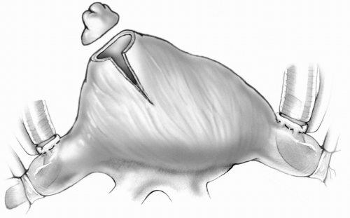
A median sternotomy is used, and standard bicaval cannulation is performed. The initial right atrial incisions and lesions are accomplished on cardiopulmonary bypass with a beating heart. After tightening the caval tourniquets, the right atrial appendage is excised. A lateral incision is made from the midportion of the base of the amputated appendage extending inferiorly 3 to 4 cm (Fig. 13-1).
The right atrium is incised beginning just anterior to the interatrial groove at the level of the right superior pulmonary vein. The incision is carried upward toward the atrioventricular groove, leaving at least 1 cm between this opening and the lateral incision from the base of the appendage (Fig. 13-2).
The irrigated radiofrequency probe is applied to the endocardial surface to create transmural lesions from the posterior aspect of the right atriotomy superiorly into the orifice of the superior vena cava and inferiorly into the inferior vena cava (Fig. 13-2). A retractor is placed under the superior edge of the atriotomy to expose the base of the amputated appendage, and a radiofrequency lesion is created from its medial aspect down to the annulus of the tricuspid valve. Another lesion is made connecting the anterior extent of atriotomy to the tricuspid valve annulus (Fig. 13-3).
If a patent foramen ovale or small atrial septal defect is present, the right atrial lesions must be performed after the aorta is cross-clamped or with induced ventricular fibrillation to prevent air embolism. The absence of a patent foramen ovale must be confirmed by transesophageal echocardiography in the operating room before instituting cardiopulmonary bypass.
The irrigated radiofrequency probe must be drawn slowly over the tissue to be ablated to achieve a transmural lesion. The thicker the atrial wall is, the longer the ablation will take. Discoloration of the endocardium should be apparent.
The opening on the base of the atrial appendage and its inferior extension are closed with 5-0 or 4-0 Prolene suture. The aortic cross-clamp is applied, and antegrade cardioplegia is given. An incision is made in the right superior pulmonary vein and extended anteriorly to meet the right atriotomy. The atrial septum is opened into the fossa ovalis (Fig. 13-4). Retractors are placed underneath the atrial septum to expose the interior of the left atrium. Lesions are created around the left and right pulmonary vein orifices, and these two lesions are connected superiorly with a straight line (Fig. 13-5). The left atrial appendage can be amputated and its base oversewn with a 4-0 Prolene double-layer suture or a radiofrequency lesion is performed around the base of the appendage. An ablation line is then drawn from the left atrial appendage to the line surrounding the left pulmonary veins (Fig. 13-5).
 FIG 13-1. Excision of the right atrial appendage and lateral incision from the base of the amputated appendage. |
Stay updated, free articles. Join our Telegram channel

Full access? Get Clinical Tree


