STEMI: Adjunctive Therapies for Primary PCI


STEMI: Adjunctive Therapies for Primary PCI


Vijay S. Ramanath MD, FACC

Craig A. Thompson MD

Julie M. Miller MD, FSCAI, FACC


In this chapter, we will discuss the following adjunctive therapies used in primary percutaneous coronary intervention (PCI) in the setting of ST-segment elevation myocardial infarction (STEMI): (a) thrombus removal devices, (b) anticoagulation, and (c) antiplatelet (parenteral and oral) agents.


THROMBECTOMY

Several clinical trials and a meta-analysis support the use of manual aspiration thrombectomy during primary PCI to improve clinical outcomes (i.e., MACE). No consistent clinical benefit or reduction in MACE has been demonstrated for routine rheolytic thrombectomy in primary PCI. Several meta-analyses have also suggested a trend to increased stroke risk in patients undergoing thrombectomy.


ASPIRATION THROMBECTOMY

This technique for STEMI patients went into widespread use, particularly after the results of the TAPAS study were published (1, 2). The TAPAS study was a single-center, randomized trial of 1,071 patients with STEMI undergoing primary PCI. Patients were randomized prior to angiography to routine manual thrombus-aspiration or conventional PCI with balloon angioplasty, followed by stenting. The study assessed angiographic and electrocardio-graphic signs of myocardial reperfusion. In patients randomized to thrombus aspiration, 55.1% underwent thrombus aspiration, followed by direct stenting; 28.6% underwent thrombus aspiration, followed by balloon angioplasty, followed by stenting; and 10.1% crossed over to standard PCI. BMS were used in 92% of procedures, and all patients were treated with aspiration, 600-mg clopidogrel, and abciximab. Approximately 57% of patients had TIMI 0-1 flow, and visible thrombus was present in ˜47% of cases. The primary endpoint, myocardial blush grade of 0 or 1, occurred in 17.1% of the patients in the thrombus-aspiration group and in 26.3% of those in the conventional PCI group (p < 0.001). (Fig. 19-1) Complete resolution of ST-segment elevation occurred in 56.6% and 44.2% of patients, respectively (p < 0.001). (Fig. 19-2) At 30 days, the rate of death in patients with a myocardial blush grade of 0 or 1, 2, and 3 was 5.2%, 2.9%, and 1.0%, respectively (p = 0.003), and the rate of adverse events was 14.1%, 8.8%, and 4.2%, respectively (p < 0.001). MACE at 30 days was not significantly different, but with important trends as follows: death (2.1% vs. 4.0%, HR: 0.52; 95% CI: 0.26-1.07; p = 0.07), reinfarction (0.8% vs. 1.9%;
HR: 0.40; 95% CI: 0.13-1.27; p = 0.11); and MACE (6.8% vs. 9.4%; HR: 0.72; 95% CI: 0.48-1.08; p = 0.12) (1).


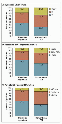
FIGURE 19-1 Myocardial reperfusion data on angiography and electrocardiography, according to treatment group.The percentages of patients are shown according to myocardial blush grade on the angiogram (Panel A) and the degree of resolution of ST-segment elevation (Panel B) and persistent ST-segment deviation (Panel C) on the electrocardiogram. PCI denotes percutaneous coronary intervention. Thrombectomy significantly improved markers of myocardial reperfusion (blush and ECG changes), with a strong 30-day trend toward a reduction in mortality. Note that 1-year mortality was significantly reduced (from 7.6% to 4.7%, p = 0.042). (From: Svilaas T, et al. N Engl J Med. 2008;358(6):557-567, with permission.)


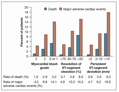
FIGURE 19-2 Rates of death and major adverse cardiac events, according to myocardial blush grade and ST-segment variables. Rates are shown for the 968 patients. p = 0.003 for the association between myocardial blush grade and death. p < 0.001 for all other associations: between myocardial blush grade and major adverse cardiac events, between ST-segment resolution and death, between ST-segment resolution and major adverse cardiac events, between persistent ST-segment deviation and death, and between persistent ST-segment deviation and major adverse cardiac events. These data support the use of surrogate markers of reperfusion. (From: Svilaas T, et al. N Engl J Med. 2008;358(6):557-567, with permission.)

The key take-home points from TAPAS were the observed improved 1-year mortality rate (4.7% vs. 7.6%) and the improved rates of 1-year cardiac mortality or recurrent nonfatal myocardial infarction (MI), among recipients of aspiration thrombectomy (2). Overall, the weight of evidence lends credence that aspiration thrombectomy protects the coronary microcirculation during primary PCI, which further explains its association with improved clinical endpoints. Still, however, because there are no data comparing selective aspiration use versus routine aspiration use (TAPAS compared routine aspiration use with no aspiration use), the 2009 update of the ACC/AHA guidelines give aspiration thrombectomy a Class IIa recommendation among STEMI patients.


RHEOLYTIC THROMBECTOMY

This form of aspiration (i.e., Angiojet) invokes the use of a catheter and high-velocity saline injection to produce a vacuum for extraction of thrombus. In doing so, it invokes fragmenting the thrombus, which may be advantageous in settings of large thrombus burden. Angiojet was studied in the AIMI study, in which 480 patients were randomized to either this device or conventional PCI (3). Importantly, the presence of thrombus was not part of the inclusion criteria in this study, and rates of baseline TIMI 3 grade flow were lower in the Angiojet cohort. Moreover, the device was activated in vivo only after it was distal to the index lesion (retrograde thrombectomy), which may have resulted in distal embolization. In the setting of these design flaws and criticisms of the trial, recipients of Angiojet were observed to have >4x increase in 30-day mortality (4.2% vs. 0.8%) and were also found to have larger infarct size. Angiojet was more recently studied in the JETSTENT trial, which was designed to correct some of the flaws in the design of the AIMI trial. This trial randomized approximately 500 STEMI patients with definite angiographic evidence of thrombus (4). MACE rates among Angiojet recipients were 11% vs. 19% among patients who underwent direct stenting (Fig. 19-3). The JETSTENT trial, given its recent publication date, was not included in the 2009 update of the ACC/AHA guidelines for STEMI care. Thus, the current ACC/AHA guidelines do not recommend the routine use of Angiojet for STEMI patients.


EMBOLIC PROTECTION

Three principal categories of devices exist: Proximal occlusive devices, distal occlusive devices, and filter-based systems. Embolic protection devices have been clearly demonstrated to be advantageous during SVG interventions. However, their effectiveness during primary PCI in native coronary arteries has not been shown in randomized clinical trials (neutral effect).

These devices are deployed downstream to the lesion (distal protection) in the hope of reducing macro- and microembolization of debris. The efficacy of such devices has been studied in a variety of clinical trials. The EMERALD trial randomized 500 STEMI patients to distal protection versus conventional PCI (5). Distal protection recipients had longer door-to-balloon times and tended to have larger infarcts. This may have contributed to the lack of difference in mortality or MACE rates at 6 months between the
two groups, despite the fact that embolic debris was removed from 73% of patients randomized to distal protection. Similar clinical outcomes were seen in the ASPARAGUS trial (6) and others investigating embolic protection devices (Table 19-1). Thus, while embolic protection may be useful in situations of high thrombus burden, where there is significant concern for distal embolization, the routine use of embolic protection is not recommended in primary PCI for STEMI patients.


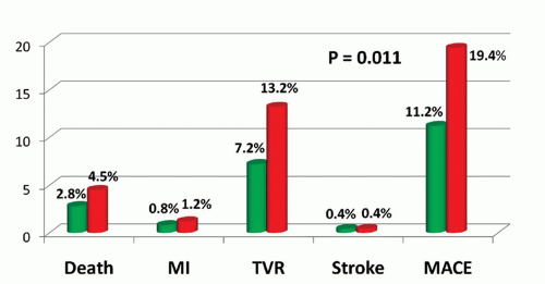
FIGURE 19-3 Clinical outcomes at 6 months of rheolytic thrombectomy versus direct stenting from the JETSTENT study. Rheolytic thrombectomy (RT) is shown in green, and direct stenting (DS) in red. MACE, major adverse cardiovascular events; MI, myocardial infarction; TVR, target vessel revascularization. (From: Migliorini A, et al. J Am Coll Cardiol. 2010;56: 1298-1306, with permission.)


TABLE 19-1 Randomized Trials of Distal Protection Devices in Primary PCI


Trial


N


Study Device


GP IIb/IIa Use, %


Primary Endpoint


Results


Outcomes


EMERALD


501


Guard Wire plus


83


STR > 70%; infarct size


63.3% vs. 61.9% (p = NS);


No difference in STR and infarct size between groups


12.0% vs. 9.5% (p = NS)


No difference in 6-month MACE


ASPARAGUS


341


Guard Wire plus


Not reported


STR > 70%


38.2% vs. 35.5% (p = NS)


Less distal embolization in treatment group


MBG > 3 ??????(30 day)


42.9% vs. 30.4% (p = 0.035)


PROMISE


200


Filter wire


100


Flow velocity in IRA


34 ± 17 vs. 36 ± 20 cm/s (p = NS)


No improvement in coronary reperfusion


PREMIAR


140


SpideRX


26


STR ≥ 70%


61.2% vs. 60.3% (p = NS)


No difference in STR or MBG


UPFLOW


100


Filter Wire-EX


75


TIMI 3 flow; STR ≥ 70%


88.2% vs. 93.9% (p = NS)


No difference in angiographic or ECG evidence of reperfusion


68.1% vs. 66% (p = NS)


65.6% vs. 64.3% (p = NS)


Reproduced from: Srinivasan, et al. Circulation. 2009; 119(9):1311-1319.


May 28, 2016 | Posted by in CARDIOLOGY | Comments Off on STEMI: Adjunctive Therapies for Primary PCI

Full access? Get Clinical Tree

Get Clinical Tree app for offline access