Elective Percutaneous Coronary Intervention for Stable Coronary Artery Disease and Silent Myocardial Ischemia


Elective Percutaneous Coronary Intervention for Stable Coronary Artery Disease and Silent Myocardial Ischemia


Ronan Margey MB, MRCPI

Douglas E. Drachman MD, FSCAI, FACC


Coronary artery disease (CAD) remains the leading cause of mortality in most industrialized countries (1). The World Health Organization estimated that 7.2 million global deaths occurred due to CAD in 2002, and that the number may exceed 11 million by 2020 as the world population continues to age (2). Despite these staggering figures, the age-standardized mortality related to CAD has fallen by more than 40% over the past two decades: half of the decline is attributed to improved primary preventive strategies, in concert with better early detection and reduction of major CAD risk factors; the other half is attributed to advances in medical and interventional therapies, particularly those related to the management of patients with acute coronary syndromes (ACSs) (3).

CAD results from the progressive formation of atherosclerotic plaque in the vessel wall (4, 5). Mechanisms such as vascular inflammation, endothelial dysfunction, intraplaque hemorrhage, and plaque rupture may contribute to endoluminal disruption, with consequent arterial thrombosis and acute occlusion (1, 6). The spectrum of clinical syndromes related to coronary atherosclerosis may be highly variable, depending on the location and degree of vessel stenosis and the potentially dynamic influence of plaque disruption and degree of vascular occlusion. Along this spectrum, CAD may be clinically inapparent, with asymptomatic (“silent”) episodes of myocardial ischemia, or may produce symptomatic ischemic syndromes, including stable reproducible myocardial ischemia (characterized by angina pectoris), unstable myocardial ischemia (unstable angina), acute myocardial infarction (MI), congestive heart failure, arrhythmia, or sudden death.

The main therapeutic objectives for patients with CAD are to relieve anginal symptoms and to prevent adverse cardiovascular events. Medical treatment strategies may reduce the biological activity within coronary plaques—so-called “plaque stabilization”— thereby reducing plaque formation and preventing future ischemic events by staving off plaque rupture. Medical therapies may also reduce myocardial oxygen and energy requirements, attenuating symptoms in the context of fixed coronary stenoses. Revascularization by either percutaneous coronary intervention (PCI) or coronary artery bypass grafting (CABG) surgery may improve myocardial perfusion in the context of flow-limiting coronary stenoses, thereby reducing ischemia and the associated clinical manifestations.

Several large-scale, randomized clinical trials have demonstrated that compared with medical therapy, an “early invasive” approach with PCI may reduce adverse cardiovascular events—including death and recurrent MI—in patients who present with unstable, ACSs (7, 8). In comparison, however, the outcomes of applying an “early invasive” approach to patients with chronic stable angina remain far less clearly defined, and remain a topic of considerable controversy (9, 10). On the one hand, it is not presently possible to identify which coronary stenoses may ultimately become “vulnerable” and cause adverse future events; and the presence of ischemia itself may confer long-term risk. On the other hand, PCI carries inherent, albeit small, risk. In chronic, stable coronary syndromes, it is difficult to calculate the “trade-off” point where the risk of performing PCI is offset by future benefit from reducing the associated ischemia or the risk that the lesion may one day become unstable.

These limitations having been noted, it remains that the majority of elective PCI procedures—more than 400,000 annually in the United States—are performed for patients who present with chronic stable angina (11). Of these patients, fewer than 10% have documentation of myocardial ischemia with noninvasive testing, and only 44% are documented to have received an adequate trial of optimal medical therapy (OMT) prior to PCI (12, 13).

Recent clinical trials and meta-analyses have demonstrated superior improvements in symptom control and quality of life with PCI compared with medical therapy in patients with chronic stable angina (14, 15). Most have failed to demonstrate improvement in survival or reduction in MI with PCI compared with medical therapy, however (16). Although the mortality associated with unstable coronary syndromes has declined in recent years, the mortality from stable CAD remains unchanged (2). Increasingly, evidence suggests that not all angiographically significant coronary stenoses cause ischemia, and that the approach of performing PCI for all angiographically significant lesions may, on balance, lead to greater adverse outcomes and the lack of benefit of PCI over medical therapy (17, 18 and 19).

In this chapter, we will review the epidemiology, pathophysiology, and prognosis of symptomatic and silent myocardial ischemia (SMI). With the perspective of contemporary practice, which emphasizes cost-effectiveness and appropriate use of medical therapies and intervention, we will provide review and future perspective on the roles of optimal medical and revascularization strategies for managing patients with stable CAD and SMI.


DEFINITION, EPIDEMIOLOGY, AND PROGNOSIS OF STABLE CAD AND SMI


Chronic Stable Angina

The diagnosis of angina pectoris is derived from the clinical history, classically described as exertional chest pain, relieved with rest or following administration of sublingual nitroglycerin. Chronic stable angina refers to the clinical syndrome in which the frequency and severity of angina is consistently provoked by a predictable amount of physical exertion or emotional stress over time (6, 10).

Of the estimated 17 million individuals in the United States with CAD, approximately 10 million report angina pectoris (2). The prevalence is higher in men and increases with age by a factor of 10-fold between the ages of 50 and 70. Chronic stable angina is the cardinal manifestation in more than half of patients newly identified to have CAD, and confers a substantially higher mortality than the average population, increasing with age (1). Populationbased data from the Framingham Heart Study, predating the widespread adoption of antiplatelet therapy, βblockers, and aggressive
risk factor modification, identified an annual mortality of 4% in patients with chronic stable angina (6).


Silent Myocardial Ischemia

Silent (asymptomatic) myocardial ischemia (SMI) is defined as objective evidence of myocardial ischemia in the absence of angina or angina equivalents (20). The clinical scenario was first described in the 1970s, and has subsequently been recognized as an important indicator of adverse prognosis (21).

SMI may be identified in individuals who develop signs of ischemia in the absence of symptoms during exercise or pharmaceutical stress testing. SMI was traditionally diagnosed using ambulatory electrocardiography (EKG) monitoring.

Cohn et al. (22) proposed a classification schema for asymptomatic myocardial ischemia: (a) Type I SMI, describing asymptomatic individuals with CAD but no history of prior MI; (b) Type II SMI, describing asymptomatic individuals with a history of prior MI; and (c) Type III SMI, describing individuals with both symptomatic and asymptomatic episodes of ischemia (23).

SMI is common in the general population, described in 3% of the overall population older than 60 years and in 10% of those over 70 (20). In one of the earliest studies of exercise testing, 1,390 men in the U.S. Air Force were evaluated: 111 had abnormal findings, of whom 34 (2.5% of the original population tested) were found to have coronary artery lesions of >50% stenosis (22, 23). Thaulow et al. evaluated 2,014 Norwegian male office workers with stress testing, and confirmed the presence of significant coronary lesions at angiography in 2.7% of the study population (24). In the Framingham Heart Study, 5,127 asymptomatic patients were followed up for 30 years, where 35% of females and 28% of males developed EKG evidence consistent with MI (20, 22). Kral et al. studied the impact of silent ischemia over 25 years in asymptomatic patients with positive family history of CAD: 28% of male siblings with a positive myocardial perfusion study (MPS) developed clinically manifest CAD compared with 12% of those with a negative MPS, with a mean time of 8 years between detection of silent ischemia to the first cardiovascular event (25).

Silent ischemia is a common finding in patients with traditional cardiac risk factors. In one study, 15% of patients with mild to moderate hypertension without symptoms or signs of CAD were found to have asymptomatic ST-segment depression during ambulatory EKG or exercise testing (26). In asymptomatic individuals with type II diabetes mellitus, 12% were identified to have abnormalities on exercise stress testing, with half of this 12% having abnormal myocardial perfusion studies (27). In individuals with diabetes plus at least one other CAD risk factor but no overt symptoms, 33% have evidence of silent ischemia.


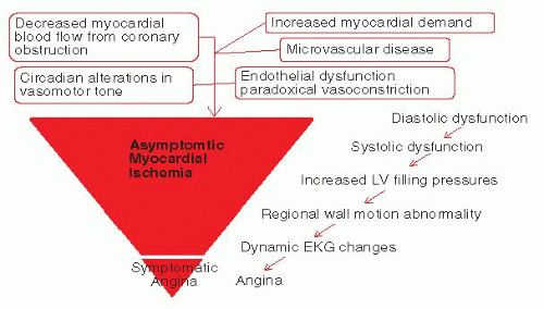
FIGURE 15-1 The iceberg effect of the ischemic cascade—the burden of asymptomatic myocardial ischemia and the tip of the iceberg—symptomatic angina.

In patients with documented CAD, episodes of silent ischemia occur frequently, despite apparent symptom control with medication. The presence of these asymptomatic episodes is associated with an increased risk of death and MI. On balance, asymptomatic ischemia occurs more frequently than symptomatic ischemia in patients with stable CAD (28, 29, 30 and 31). Figure 15-1 highlights the ischemic cascade that results from the reduction of coronary blood flow because of coronary stenosis, or the relative imbalance of myocardial oxygen demand compared with delivery in the context of flow-limiting stenosis. The diagram highlights the concept that symptoms of angina represent the final manifestation of ischemia, with substantial asymptomatic hemodynamic and electromechanical consequences occurring well before the onset of chest pain. Using ambulatory EKG monitoring, 50% of individuals with stable CAD were found to have asymptomatic ST-segment depression. Additionally, more than 50% of patients monitored with telemetry during admission for unstable angina are found to have asymptomatic episodes of ischemic ST-segment changes. Sudden cardiac death comprises 18% of all primary clinical presentations with CAD; and more than 50% of sudden deaths occur without an antecedent history of CAD. As many as 40% of patients with stable angina treated with one or more antianginal medications, and 30% to 40% of patients after MI, have episodes of asymptomatic ischemia (20, 22, 29).

The presence of asymptomatic ischemia confers an elevated risk of adverse cardiovascular events. In the Multiple Risk Factor Intervention Trial (MRFIT) of over 12,000 asymptomatic middle-aged men with two or more CAD risk factors, the presence of ischemia during exercise testing was highly predictive of future cardiac death (relative risk [RR]: 3.4) (23, 32). In the Lipid Research Clinic Primary Prevention Trial (LRCPPT) of greater than 6,000 males without prior CAD, asymptomatic ischemia on submaximal exercise testing was associated with a significantly greater age-adjusted cardiovascular mortality (33). In patients with established CAD identified at an index presentation with MI or unstable angina, 30% to 40% are subsequently found to have evidence for SMI, which is associated with a higher rate of future cardiovascular events and death (30).


The presence of SMI also confers a higher risk of adverse cardiovascular events in patients with documented CAD and chronic stable angina. In the Asymptomatic Cardiac Ischemia Pilot (ACIP) Study, the 1-year composite rate of death, MI, and hospital admission was 13% in 558 patients with SMI (34). The Coronary Artery Surgery Study (CASS) included 880 patients with documented CAD, and found that the 7-year incidence of MI and death was greatest in those with asymptomatic ischemia on exercise testing (26%) compared with 23% in patients with symptomatic ischemia on exercise testing and 2% in patients with no demonstrable ischemia (35).

More recent studies suggest that the identification of SMI during ambulatory EKG monitoring is a stronger predictor of future adverse events than ischemia found on exercise testing. In one study, 12.5-month follow-up of 86 patients with stable CAD and ischemia on exercise testing found a correlation between the number of ST-segment depression events on ambulatory EKG monitoring with the duration of exercise, time to onset of ST-segment depression, and the depth of ST-segment depression at stress testing. Following multivariate adjustment, however, only ST-segment depression on ambulatory monitoring significantly predicted adverse future events. In another study, the presence of SMI during ambulatory monitoring was a more powerful predictor of mortality than exercise duration, age, prior MI, hypertension, diabetes, or smoking status in asymptomatic patients with CAD on antianginal therapy (20, 28, 30, 31). The most important factors in determining outcome in patients with CAD are the presence and extent of ischemia (36, 37).


PATHOPHYSIOLOGY OF SYMPTOMATIC ANGINA PECTORIS AND SMI

Myocardial ischemia is caused by an imbalance between myocardial oxygen requirements or demands and myocardial oxygen supply (5). The most important determinants of myocardial oxygen demand are heart rate, wall stress or tension, and contractility, which are influenced by activity and sympathetic tone. Myocardial oxygen supply is primarily increased by augmenting coronary blood flow.

When coronary stenosis is severe, coronary blood flow may be limited, particularly at times of increased demand, and results in ischemia. In the presence of vascular inflammation and endothelial dysfunction, exercise may provoke paradoxical vasoconstriction, rather than vasodilatation of the coronary arteries, further reducing myocardial perfusion. In cases of critical stenosis, coronary blood flow and myocardial oxygen supply may be so reduced as to cause ischemia even at rest or with vasoconstriction from elevated sympathetic tone. At a microvascular and cellular level, adaptive mechanisms, such as intracellular signaling with adenosine, increase myocardial capillary dilatation, reduce flow resistance, and optimize oxygen extraction. Because coronary blood flow is maximal in diastole, the perfusion gradient across the myocardium may be influenced by changes in wall tension and left ventricular filling pressures. The left ventricular wall tension is greatest at the subendocardial surface, which therefore is typically the first site of ischemia (5, 22).

The precise mechanisms that determine when myocardial ischemia is silent or symptomatic remain to be elucidated. Ambulatory EKG monitoring demonstrates the propensity for asymptomatic ischemia in the morning, suggesting that a circadian pattern of increased myocardial oxygen demand related to increased heart rate and blood pressure may provoke some asymptomatic ischemia. Circadian rhythms may also alter vasomotor tone, platelet activity, and in vivo fibrinolytic activity, with implications on myocardial perfusion (38, 39). Heterogeneity in peripheral and central nervous neural processing and nociception has also been implicated in patients with asymptomatic versus those with symptomatic ischemia. The presence of autonomic neuropathy and increased endogenous endorphin levels have been proposed as mechanisms that enable episodes of asymptomatic ischemia (20, 22).

At the most fundamental level, myocardial ischemia reflects the mismatch between supply of and demand for blood flow and oxygen, resulting in a stereotyped sequence of hemodynamic and electromechanical alterations, the final step of which is the development of symptomatic angina. Figure 15-1 describes this ischemic cascade, and highlights the numerous subclinical alterations that occur before the onset of chest pain. When coronary arterial supply is outstripped by demand, first diastolic, then systolic regional wall dysfunction results. As a consequence, left ventricle (LV) filling pressures rise, worsening the regional wall abnormality. Following mechanical disruption of LV function, the EKG becomes abnormal; and, only at this point, will the patient begin to develop symptomatic angina. This physiologic cascade reflects the “iceberg” concept of symptomatic and asymptomatic angina: the ultimate manifestation of angina pectoris represents only the final culmination of events, or the tip of the iceberg, while the majority of ischemic manifestations occur silently, beneath the surface of clinical detection.


MANAGEMENT OF STABLE CAD AND SMI

The main goals in managing patients with stable CAD include the relief of symptoms, resulting in freedom from angina and improved quality of life, and the reduction of future cardiovascular events and mortality, through coronary plaque stabilization and slowing the progression of atherosclerosis. Regardless of patient symptomatology, the reduction of residual ischemia is the most important determinant of future prognosis and outcomes.

Comprehensive management of stable CAD requires a multifaceted simultaneous approach;


  • 1. Identify and treat any associated medical conditions that may worsen or precipitate angina, such as thyrotoxicosis or anemia.


  • 2. Modify established CAD risk factors and commence secondary preventative medications.


  • 3. Modify lifestyle factors.


  • 4. Commence, titrate, and ensure compliance with antianginal pharmacotherapy.


  • 5. Perform revascularization (PCI or CABG) for persistent symptoms, or substantial residual ischemia on medical therapy.

The above treatment strategies are further outlined in Table 15-1. The current American College of Cardiology/American Heart Association recommendations for pharmacotherapy of stable CAD are outlined in Table 15-2, and the lifestyle goals are outlined in detail in Table 15-3 (9, 10).

Over the past three decades, significant advances in the array of medications targeting secondary prevention and symptomatic treatment of CAD have resulted in dramatic improvements in survival among patients with stable CAD (3). In the Framingham heart study, prior to the widespread adoption of current medical therapy, the annual mortality from stable CAD was 4% (2).
In comparison, patients receiving contemporary medical therapy for stable CAD (92% were taking platelet inhibitors, 62% βblockers, and 58% lipid-lowering therapy) were evaluated in the EUROPA trial and had an annual risk of cardiovascular death or MI of 2.5% (40). Of the medical therapies available, aspirin, angiotensin converting enzyme (ACE) inhibitors, and lipid-lowering statin medications have been proven to reduce mortality and morbidity in patients with stable CAD and preserved left ventricular function. To avoid one death or MI, about 175 patients need to be treated with aspirin for 1 year (relative risk reduction: 23%); 120 patients with standard dose statin medications (relative risk reduction: 30%); and 200 patients with an ACE inhibitor (relative risk reduction: 20%) (1). The other medications, including longacting nitrates, βblockers, and calcium-channel antagonists, have been shown to improve symptomatology, exercise tolerance, and quality of life among patients with stable CAD; but their effect on survival has not been definitely established, with the exception of βblockers in patients with stable CAD and impaired left ventricular function (1, 10).


TABLE 15-1 Medical Therapies for Chronic Stable Angina


Management Strategy


Intervention


1. Treatment of associated conditions that


1. Check for anemia


may precipitate or worsen angina


2. Check for thyrotoxicosis


3. Control tachyarrhythmias


4. Check for concomitant left ventricular failure or valvular heart disease


5. Check for cocaine use


2. Reduction of coronary risk factors


1. Blood pressure control


2. Smoking cessation


3. LDL cholesterol reduction


4. HDL cholesterol elevation


5. Diabetes control


6. Physical exercise


7. Inflammation reduction


8. ACE inhibitors


3. Lifestyle adjustments


1. Weight loss


2. Physical exercise


3. Stress reduction


4. Antioxidants and dietary supplements


4. Antianginal Pharmacotherapy


1. Antiplatelet therapy


2. βblocker therapy


3. Combination of βblocker with calcium-channel antagonist or long-acting nitrates


4. Ranolazine


5. Nicorandi


6. Ivabradine


7. Fasudi


8. Metabolic agents-trimetazidine


5. Revascularization


1. Percutaneous coronary intervention (stenting)


2. Coronary artery bypass grafting


A detailed discussion of the pharmacodynamics, side effects, interactions, and contraindications of each of the above medication groups is beyond the scope of this chapter. We will briefly outline the evidence supporting the antianginal efficacy of each of the major medication groups. Long-acting nitrates are highly effective antianginal agents, reducing the frequency and duration of ischemic episodes and producing total suppression of ischemia in 35% of patients (1, 20). Side effects and medication intolerance, most commonly, headaches, must be considered. β blockers represent the cornerstone of antianginal therapy, reducing myocardial oxygen demand, heart rate with exercise, resting heart rate, blood pressure, circadian effects, and ventricular contractility. In pooled analyses, βblockers reduce the frequency and duration of silent ischemic episodes by 59% and 69%, respectively, with total abolition of ischemia in 55% of patients. βblockers are contra-indicated in patients with severe asthma or significant conduction system disease. Calciumchannel antagonists may be used as an alternative to βblockers for patients with contraindications or side effects. In pooled analyses, the use of calcium-channel blockers results in 46% reduction in
frequency and a 36% reduction in duration of ischemic episodes (1, 22, 41). Prior studies have examined the use of antianginal medications effecting combination for potential synergy. In the ACIP study, the combination ofβblocker and calcium-channel antagonist resulted in total ischemia suppression of 48% compared with the combination of calcium-channel antagonist and long-acting nitrate (total suppression 33%) (34). Current treatment guidelines recommend initial therapy with an antiplatelet agent and β blocker, with subsequent addition of a long-acting nitrate if symptoms persist. Calcium-channel antagonists are most commonly used in place of β blockers, and addition of a second agent is recommended if symptoms persist. Numerous additional antianginal agents are available for use, but a detailed discussion of these agents is beyond the scope of this chapter. They are listed in Table 15-1.


TABLE 15-2 ACC/AHA Recommended Pharmacotherapy for Chronic Stable Angina


Class


Indication


Level of Evidence


I (indicated)


1.


Aspirin in the absence of C/l


A


2.


βblockers as initial therapy in the absence of C/l in all patients with or without prior Ml


A, B


3.


ACE inhibitor in all patients with impaired LV systolic function, chronic kidney disease and/or diabetes


A


4.


LDL reduction therapy in patients with documented or suspected CAD and LDL >130 mg/ dL, goal LDL<100 mg/dL, or <70 mg/dL if prior Ml


A


5.


Sublingual nitroglycerin for immediate relief of angina


B


6.


Calcium-channel antagonists or long-acting nitrates as initial therapy when βblockers C/l


B


7.


Calcium-channel antagonists or long-acting nitrates in combination with βblockers for persistent symptoms despite βblocker titration (avoid short acting dihydropyridine calcium-channel antagonists)


B


8.


Calcium-channel antagonists and long-acting nitrates in combination as a substitute for β blockers if βblocker therapy produces side effects


C


lla (good evidence)


1.


Clopidogrel when aspirin is absolutely C/l


B


2.


Long-acting nondihydropyridine calcium antagonists instead of βblockers as initial therapy


B


3.


In patients with documented or suspected CAD and LDL cholesterol 100-129 mg/dl; lifestyle or drug therapy to lower LDL <100 mg/dL or <70 mg/dL if prior Ml; weight reduction and increased physical activity in persons with metabolic syndrome; and initiation of therapy to lower triglycerides or raise HDL cholesterol if appropriate


B


4.


ACE inhibitor in patients with CAD or other vascular disease


B


llb (weak evidence)


1.


Low intensity anticoagulation with warfarin in addition to aspirin


B


lll (not indicated)


1.


Dipyridamole


B


2.


Chelation therapy


B


TABLE 15-3 Specific lifestyle goals in patients with chronic stable angina


Risk Factor/Strategy


Goal


Smoking


Complete cessation, counseling and medications if necessary


Blood Pressure


<140/90 or <130/80 if CHF, chronic kidney disease, or diabetes


Lipid management


LDL <100 mg/dL; or less than 70 mg/dL if prior Ml, stroke, or diabetes


Physical Activity


Minimum 30 minutes, 3-4 times weekly. Daily 30-minute exercise optimal


Weight Management


BMI: 18.5-24.9 kg/m2


Diabetes


HbA1C as near normal as possible; <7% minimum


C/l, contra-indication; Ml, myocardial infarction; ACE, angiotensin converting enzyme; LV, left ventricle; CAD, coronary artery disease; LDL, low-density lipoprotein; HDL, high-density lipoprotein; CHF, congestive heart failure; BMI, body mass index; HbA1c, Glycosylated hemoglobin percentage


WHAT CONSTITUTES OMT?

There is no uniform definition of OMT. OMT for stable CAD has evolved significantly over the last 30 years. Societal guidelines for secondary prevention therapy were not introduced until 1995,
with most therapy prior to this targeted toward ACS patients. The earlier trials of medical therapy for stable angina used different drug preparations and combinations, and different drug dosages and titration schedules (34). The sole lifestyle modification targeted was smoking cessation. It was not until the early 2000s that a greater emphasis was placed on β blocker therapy and ACE inhibitor use in most patients with coronary heart disease (10, 40). Successive treatment guidelines have broadened the indications for cholesterol reduction therapy, with endorsement of progressively earlier initiation of statin therapy and lower lowdensity lipoprotein (LDL) targets. Similarly, targets for blood pressure control have been serially reduced. Over time, a more significant emphasis has been placed on diet, exercise, weight control, diabetes management, and blood pressure control. In light of the evolution of medical therapy over time, it is difficult to compare the findings of multiple studies of medical therapy versus revascularization from different eras, or even to extrapolate the findings of prior studies to current practice, as therapeutic options, targets, and management strategies have changed so dramatically.

Tables 15-2 and 15-3 outline the latest ACC/AHA treatment guidelines for chronic stable angina (10). Despite the publication of these guidelines, however, prescribing rates of “OMT” and patient compliance with medical therapy remain poor. In 2011, Borden et al. reported an analysis of the patterns and intensity of OMT in patients undergoing PCI from the National Cardiovascular Data Registry (NCDR), demonstrating that fewer than half of patients undergoing PCI for stable CAD were receiving OMT (11

Only gold members can continue reading. Log In or Register to continue

Stay updated, free articles. Join our Telegram channel

May 28, 2016 | Posted by in CARDIOLOGY | Comments Off on Elective Percutaneous Coronary Intervention for Stable Coronary Artery Disease and Silent Myocardial Ischemia

Full access? Get Clinical Tree

Get Clinical Tree app for offline access