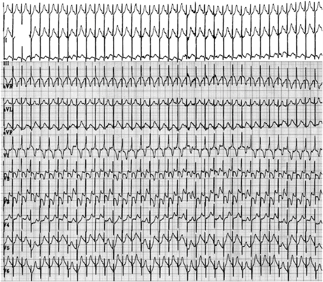
Fig. 7.1
Six (limb) lead electrocardiogram (top three and bottom three tracings) with an esophageal bipolar lead recording (middle tracing), all at paper speed of 100 mm/s, in a 2-month-old boy with tricuspid atresia. Panel A on the left demonstrates supraventricular tachycardia at 270 bpm. Panel B demonstrates sinus rhythm at 160 bpm. Note the PR interval and P-wave morphology and axis during the tachycardia are virtually identical to the PR and P-wave morphology and axis observed during sinus rhythm, suggesting the origin of the tachycardia to be around the sinoatrial (sinus) node

Fig. 7.2
Transesophageal atrial overdrive pacing (stimulus rate -s- at a pacing cycle length of 220 ms) in the patient from Fig. 7.1. Successful cardioversion to sinus rhythm is achieved, supporting the diagnosis of a reentrant mechanism in the region of the sinus node (inferred from Fig. 7.1), i.e., sinoatrial reentrant tachycardia

Fig. 7.3
Sinus tachycardia at 250 bpm in a 14-month-old girl with a urinary tract infection and fever of 40 °C
Table 7.1
Criteria for diagnosis of sinoatrial reentrant tachycardia
Sinus-like P-waves (positive P waves in limb leads 1, 2, 3, and negative P-wave in aVR) during the tachycardia |
Initiation (and resetting and termination) by appropriately timed single premature atrial (or sinus) stimuli, over an echo zone either spontaneous or through programmed extrasimulation independent of atrioventricular nodal conduction slowing (Fig. 7.1) |
Normal PR interval (identical to that during sinus rhythm) with a long RP interval during the tachycardia (Fig. 7.2) |
Termination of the tachycardia by vagal maneuvers or adenosine |
Exclusion of other mechanisms |
When considering the diagnosis of this tachycardia, one must exclude other physiologic or pathologic states that can cause a sustained acceleration of sinus rhythm. It is very unusual for the sinus rate to exceed 220 bpm under any physiologic or pathologic state; the maximal heart rate of an elite athlete is usually approximately 220 bpm under maximal sustained exercise. A good formula for the maximal attainable heart rate is: 208 bpm minus 0.7 times the subject’s age.
Hyperthyroidism, febrile illnesses, and hypovolemic states can increase the heart rate (Fig. 7.3) above what the apparent metabolic needs of the patient are at the time. Hyperthyroidism may present with an accelerated sustained heart rate usually about 120 bpm in the absence of other signs or symptoms. Febrile illnesses or hypovolemic states rarely exceed the maximal heart rate appropriate for the patient’s age.
< div class='tao-gold-member'>
Only gold members can continue reading. Log In or Register to continue


