Electrocardiographic manifestations of sick sinus syndrome
Sinus bradycardia
Extreme sinus arrhythmia
Tachycardia–bradycardia syndrome
Sinoatrial exit block
Sinus pause/sinus arrest
Sinoatrial reentrant tachycardia
Electrocardiographic findings associated with sick sinus syndrome
Intra-atrial reentrant tachycardia (IART)
Atrial fibrillation
AV nodal dysfunction
Etiology
Conditions associated with a high prevalence of sick sinus syndrome include atrial repair of d-transposition of the great arteries, complete repair for anomalous pulmonary venous drainage, atrial septal defect, and single ventricle palliation with the Fontan operation, although almost any open heart repair may result in sinus node impairment. Sinus node malformation is associated with left atrial isomerism and sinus node dysfunction may coexist with congenital complete heart block. Congenital sinus node dysfunction due to SCN5a mutation and perhaps mutations in other cardiac genes (e.g., the MYH6 gene encoding the cardiac aloha heavy chain subunit of myosin) along with congenital central hypoventilation syndrome with sinus node dysfunction are rare but have been reported. Familial clustering has been reported in the absence of structural heart disease. Extrinsic causes of sinus node dysfunction include autonomic imbalance and medications. Heavily conditioned athletes, and even normal preadolescent children (4–10 years old), may have bradycardia and sinus pauses of greater than 2 s due to prominent vagal influence. Recently, sleep apnea has been related to sinus node dysfunction. Many anti-arrhythmia medications can further impair the sinus node function particularly for patients with preexisting abnormalities. Neuromuscular disorders, such as Kearns Sayre (ophthalmoplegia, pigmentary degeneration of the retina, and cardiomyopathy), Friedreich’s Ataxia and myotonic dystrophy may have a predilection for the conduction system and sinus node in particular. Immunologic basis for sinus node dysfunction have been seen in patients with anti-sinus node (ASN) antibodies as well as in donor hearts after orthotopic heart transplantation. Most commonly anti-Ro/SS-A or anti-LA/SS-B antibodies are well established in the mechanism of congenital AV block in infants of mothers with Sjögren’s, lupus, or other undifferentiated autoimmune syndrome, but there also have been rare reports of sinus node dysfunction. While comprehensive this list may not include every cause of sick sinus syndrome.
Symptoms
Although the designation “syndrome” usually implies symptoms as part of the clinical picture, it is often difficult to elicit in the young patient. In the more extreme cases, symptoms may include pre- or frank syncope, shortness of breath, lightheadedness with activity, fatigue, weakness, and inability to do what one used to be able to.
ECG Characteristics
The ECG characteristics of sick sinus syndrome were first fully described by Ferrer, initially in the elderly. The ECG features are variable and can change within a single patient. Not all patients will manifest all of the electrocardiographic findings outlined below. Potential sources of electrocardiographic data include standard 12–15 lead electrocardiograms, Holter monitoring, event recorders, and rhythm strips from physiologic monitors. Clear documentation is not always present in short-term recordings. Multiple leads should be recorded to detail P wave morphology.
Long-term recordings with Holter provide an overview of overall heart rate variation (Fig. 14.1) and the prevalence of abnormally slow and fast rhythms. Ideally three orthogonal leads are recorded to optimize P wave recognition (i.e., Lead I, Lead II or III, and a precordial lead). Event recorders, particularly those with continuous storage capabilities, allow for correlation of symptoms to electrocardiographic changes. Newer devices have programmable parameters for automated recording of tachycardia and bradycardia episodes. An implantable device is available for special circumstances that preclude wearing an external monitor, but because it requires anesthesia and surgery for placement, it should be reserved for the patients with a diagnostic dilemma as well as high risk. Exercise testing is useful to assess the chronotropic response. All children and adolescents should be able to attain a heart rate of 180 beats per minute (near 70 % max predicted heart rates, which needs to be teased from poor effort). Patients with sinus node dysfunction also may have rate instability with exercise or exaggerated slowing of heart rate or pauses in the recovery period.
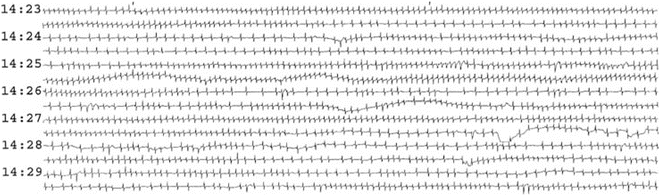
Fig. 14.1
Full disclosure printout from a Holter monitor. Individual complexes are too small for detailed evaluation, but abnormal pattern of heart rate variation is apparent
Sinus Bradycardia
Bradycardia is the most common feature of sick sinus syndrome. Bradycardia may be sustained or paroxysmal. Escape rhythms (Fig. 14.2) arise from the atrium, atrioventricular node, and ventricles, but these tissues are often dysfunctional and the escape rates are frequently lower than expected for age. Healthy children may have brief periods of sinus bradycardia with atrial or junctional escape rhythm in the absence of any sinus node pathology. The normal heart rate is age dependent. The lowest values observed during Holter recording are less than those from ECG recording. Recording artifacts and other rhythm abnormalities may confound the interpretation. An abnormally low heart rate may be due to second- or third-degree atrioventricular block as well as blocked atrial and junctional extrasystoles. A sudden loss of signal may appear similar to a sinus pause. There are often clues to this type of artifact, including resumption of recording with a T wave or a different time of drop out on another channel (Fig. 14.3). Older Holter recorders using a tape drive mechanism can suddenly speed or slow mimicking bradycardia or tachycardia. With this type of artifact, all components of the recording are expanded or compressed (Fig. 14.4). While digitalized Holter monitors will not demonstrate this kind of artifact, one could manually manipulate the ECG paper strip that is recording the rhythm and render it abnormal.
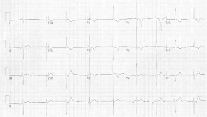
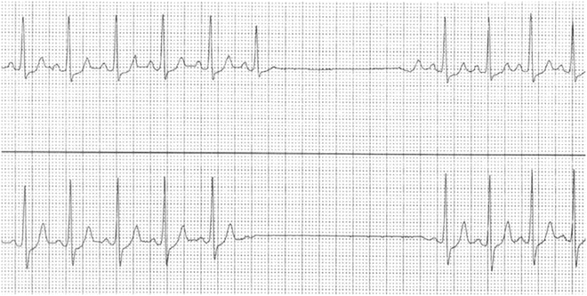
Fig. 14.2
Sinus bradycardia with alternating atrial and junctional escape rhythms in a patient 15 years after Mustard repair of d-transposition of the great arteries
Fig. 14.3
Simultaneous lead I (upper panel) and lead III (lower panel) from Holter recording. Apparent sinus pause is actually due to artifact. Signal loss is not simultaneous on both channels and lead I resumes rhythm with a T wave which is not possible
Fig. 14.4
Simultaneous lead V1 (upper) and V5 (lower) from Holter recording. Apparent bradycardia and tachycardia due to artifact from changing tape speed. Note that all components of the rhythm (P wave, QRS complex, and T wave) are compressed with tachycardia
Extreme Sinus Arrhythmia
Sinus arrhythmia is a normal pattern of heart rate acceleration and deceleration present in most children. A significant component of this variation is due to respiratory influences on the autonomic nervous system. Abnormal or extreme sinus arrhythmia is defined as greater than 100 % variation in PP intervals. Sinus node exit block may also cause variation of this magnitude.
Tachycardia–Bradycardia
The tachycardia–bradycardia syndrome is diagnosed in the presence of recurrent prolonged pauses or sustained bradycardia following paroxysms of tachycardia. The ectopic impulses of the tachycardia penetrate the sinus node and cause exaggerated overdrive suppression of already impaired automaticity, exaggerating the bradycardia (Fig. 14.5). This phenomenon is not infrequently observed acutely following overdrive pacing or DC cardioversion of atrial flutter or intra-atrial reentry tachycardia (Fig. 14.6).
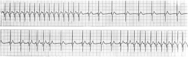
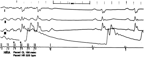
Fig. 14.5
Tachycardia–bradycardia with junctional escape in a 2-year-old
Fig. 14.6
Overdrive pacing of atrial flutter in 9-month-old girl. Note the long sinus recover time (1,300 ms) (the first beat after termination of pacing is the last reentry flutter beat) indicating suppression of the sinus node by the prolonged flutter episodes
Sinoatrial Exit Block
Sinoatrial exit block is failure of an impulse generated in the sinus node to propagate normally into the atrium. The degree of block is classified similar to the classification for atrioventricular node. A direct recording of the sinus node electrogram is necessary to diagnose first-degree sinoatrial block. The pattern of PP intervals prior to a pause can be used to infer second-degree block. Mobitz I sinoatrial block (Fig. 14.7) is manifest by a gradual shortening of PP intervals followed by a pause of less than two times the resting cycle length. Mobitz II is presumed to be the mechanism when a sudden pause of two times the resting cycle length is encountered (Fig. 14.8). Both of these are difficult to recognize in the presence of sinus arrhythmia and both patterns may be due to abnormal impulse formation in the sinus node. Neither of these conduction abnormalities can be absolutely proven without direct recording of the sinus node electrogram. Complete or third-degree sinoatrial block is indistinguishable from sinus node arrest on the surface electrocardiogram.
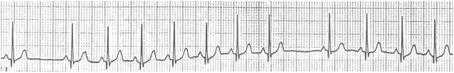
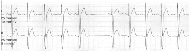
Fig. 14.7
Lead I recording suggestive of Mobitz I type sinoatrial exit block. There is a gradual shortening of PP intervals, with a consistent P wave morphology, followed by a pause of less than two times the resting cycle length. Validation of this phenomenon requires invasive recording of the sinus node electrogram
Fig. 14.8
Simultaneous lead I and II recording suggestive of Mobitz II type sinoatrial exit block. There is a pause with a PP interval that is twice the resting cycle length. Validation of this phenomenon requires invasive recording of the sinus node electrogram
Sinus Pauses and Sinus Arrest
Prolonged pauses are often present due to either sinoatrial node exit block or sinus arrest (Fig. 14.9). Sinus pauses of up to 2 s are normal in young children and adolescents. A non-sinus rhythm at a rate lower than expected for sinus rhythm is referred to as an escape rhythm and is suggestive of sinus node arrest. It too is usually normal in children without heart disease; it does not per se suggest heart disease.
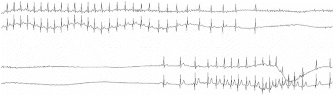
Fig. 14.9
Sinus arrest during a breath holding spell. This represents transient sinus node dysfunction due to a sudden increase in parasympathetic activity
Sinoatrial Node Reentrant Tachycardia
Sinoatrial reentrant tachycardia is uncommon. It results from abnormal conduction and reentry (see Chap. 7) within the sinus node or immediate perinodal tissue. The P waves are identical to those seen with sinus rhythm, but suggested by a sudden paroxysmal increase and decrease in rate. This rhythm cannot be distinguished from a focal atrial tachycardia in close proximity to the sinoatrial node.
< div class='tao-gold-member'>
Only gold members can continue reading. Log In or Register to continue
Stay updated, free articles. Join our Telegram channel

Full access? Get Clinical Tree


