Fig. 20.1
M-mode echocardiogram from fetus in sinus rhythm. Upper image demonstrates beam position through the atrium and ventricle. Small arrows mark atrial contractions, and arrowheads mark ventricular contraction. Note 1:1 ratio of atrial to ventricular contractions
Many factors influence the quality of the M-mode tracing. First, alignment with both fetal heart chambers can be challenging, and several different probe positions and angles should be attempted to optimize the tracing. Second, a clearer tracing will be obtained where chamber walls have the greatest excursion with contraction, such as the atrial appendage, or lateral ventricular wall.
Doppler echocardiography utilizes spectral blood flow patterns during systole and diastole as representation of atrial and ventricular contraction. The standard method is to obtain an apical four-chamber image of the fetal heart, with the pulsed Doppler cursor positioned with the gate spanning the mitral inflow as well as the aortic outflow (Fig. 20.2). The mitral “A” wave represents atrial contraction, and the systolic aortic wave represents ventricular contraction. An alternate spectral Doppler approach is to position the pulsed Doppler cursor across the SVC and ascending aorta simultaneously (Fig. 20.4a). Brief reversal of flow in the SVC during atrial systole denotes atrial contraction, with the forward systolic flow wave in the aorta showing ventricular systole. From any of these methods, the mechanical PR interval can be measured from the onset of the atrial contraction to the onset of ventricular contraction, however depicted.
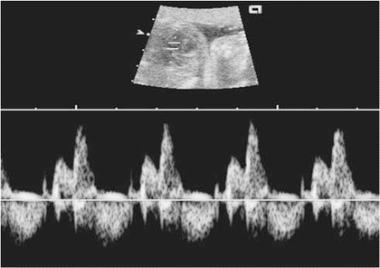
Fig. 20.2
Doppler echocardiogram from fetus in sinus rhythm. Upper image demonstrates beam position in the ventricle. Note 1:1 relationship of normal mitral inflow (E- and A-waves above baseline) and normal aortic outflow (below baseline)
New techniques including tissue velocity imaging, with creation of a “fetal kinetocardiogram,” as well as strain rate imaging have recently been described. Tissue velocity and strain rate imaging may significantly enhance current fetal echocardiographic diagnostic capabilities.
In addition to determination of the arrhythmia mechanism, echocardiographic evaluation of the fetus should incorporate assessment for any hemodynamic and anatomic abnormality. Both tachyarrhythmias and bradyarrhythmias can cause fetal heart failure, and ultimately hydrops fetalis. Complete evaluation for fetal heart failure includes assessment of heart size, ventricular systolic function, atrioventricular valve incompetence, venous Doppler patterns (increased reversal with atrial contraction) including hepatic vein, ductus venosus and umbilical venous Doppler (Fig. 20.3), and documentation of presence and size of pleural, pericardial, and abdominal effusions. All of these indices require ongoing assessment for progression by serial echocardiographic studies as well as obstetrical evaluation of fetal well-being during the mother’s pregnancy.
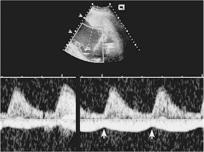
Fig. 20.3
Pulsed spectral Doppler tracing from the umbilical vein in a fetus with hydrops fetalis. Upper panel demonstrates gate position in the umbilical vein. Arrows indicate pulsations with atrial systole, consistent with high atrial pressure
Specific Fetal Arrhythmias
Extrasystoles
Extrasystoles account for 60–90 % of fetal arrhythmia. Ectopic beats may arise in the atria, junctional tissue, or ventricle. Supraventricular ectopy (SVE) is most common. Ventricular ectopy (VE) comprises fewer than 10 % of fetal extrasystole. Frequency of ectopic beats in the normal fetus has not been well established; however, the rate of extrasystoles in healthy premature infants is 20–30 %, with a slightly lower frequency in term infants.
Ectopic beats present as an irregular FHR. As in older patients, very early occurring SVE results in blocked atrioventricular conduction, either complete or partial (in the form of bundle branch block with aberrant depolarization). Hence, SVE can present as bradycardia. The majority of fetuses with premature beats are healthy, and the ectopy resolves over time.
Although the vast majority of fetuses with extrasystoles have a structurally normal heart, SVE and VE can be associated with anatomic congenital heart disease, cardiac tumors, and fetal genetic abnormalities such as Trisomy 18. In addition, SVE precedes supraventricular tachycardia (SVT) in a number of cases. Thus, all fetuses with frequent premature beats should have more frequent FHR monitoring until delivery, or until resolution of the ectopy has been sustained.
Supraventricular Ectopy
Premature atrial and junctional depolarizations occur most often as single beats. Diagnosis is made with a combination of Doppler and M-mode echocardiography. With M-mode, simultaneous atrial and ventricular recording is performed, demonstrating a normal sequence of A–V contractions and an early atrial contraction wave. The atrial beat after the premature contraction demonstrates an incomplete compensatory pause.
Doppler sampling during normal rhythm displays at the junction of the mitral inflow and left ventricular outflow tract an E-wave (early, rapid ventricular filling) followed by an A-wave (atrial contraction with active ventricular filling) and ventricular systole (with semilunar valve outflow). SVE will cause early active ventricular filling (A-wave), obscuring part of the early ventricular filling (E-wave) tracing with subsequent early ventricular systole (Fig. 20.4a). In cases of fully blocked SVE, no ventricular systole will follow the premature atrial contraction. The post-extrasystolic contraction will demonstrate a prolonged filling (diastolic) time interval. Doppler sampling of the SVC/ascending aorta will demonstrate an early flow reversal wave in the IVC during early atrial contraction, and lack of systolic wave in the aorta with a blocked SVE (Fig. 20.4b).
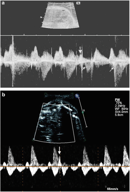
Fig. 20.4
Doppler echocardiogram from fetuses with SVE. (a) Doppler cursor through mitral inflow and aortic outflow. The large arrow marks the premature atrial contraction. The first small arrow marks the diminished aortic outflow volume with the SVE, and the second small arrow shows the increased aortic outflow volume during the post-extrasystolic contraction. Note the prolonged diastolic time interval following the SVE. (b) Doppler cursor through SVC and ascending aorta with blocked SVE. Note the atrial contractions represented by reversal in SVC above the baseline. The large arrow marks the premature atrial contraction, with no aortic outflow following the blocked SVE
The fetal PR interval can be measured using a gated pulsed Doppler technique. Premature atrial contractions should demonstrate a normal or prolonged PR interval (if slow conduction occurs, Fig. 20.5).
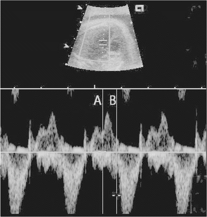
Fig. 20.5
Pulsed spectral Doppler through the mitral inflow and aortic outflow in a normal fetus, demonstrating mechanical PR interval measurement. E- and A-wave components of mitral inflow are above the baseline, whereas the aortic outflow signal is below the baseline. Calipers measure from the beginning of the A-wave (line A) to the beginning of the aortic outflow signal (line B)
The prognosis is favorable for the majority of fetuses with SVE. In addition to the conditions mentioned earlier, fetal SVE can be associated with maternal drug use or hyperthyroidism. For patients with associated maternal disease, structural congenital heart disease, tumors, or sustained tachyarrhythmias, the prognosis correlates with the associated condition. No treatment is indicated for isolated fetal SVE.
Ventricular Ectopy
M-mode echocardiography may demonstrate subtle distortion of normal ventricular contraction due to aberrant muscle conduction (Fig. 20.6). Also, a complete atrial compensatory pause is seen after most premature ventricular depolarizations. Doppler ultrasound demonstrates a characteristic AV valve inflow pattern with decreased diastolic antegrade flow. Marked retrograde flow in the IVC is seen during atrial contraction.
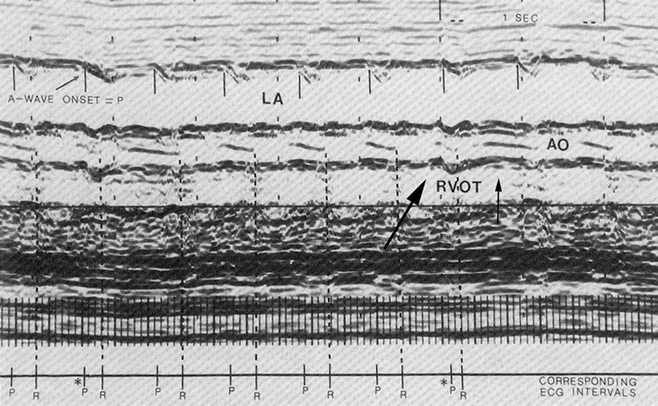
Fig. 20.6
M-mode recording from a 36-week fetus with its back toward the transducer presenting an inverted view of the heart. Premature aortic valve opening (large arrow) can be clearly seen to be followed by atrial wall contraction but with no variation in P–P interval (A-wave interval), giving a compensatory pause (small arrow, allowing diagnosis of ventricular premature beat). LA left atrium, AO aorta, RVOT right ventricular outflow tract (5 MHz transducer) [Reprinted from Crowley DC, Dick M, Rosenthal A, et al. Two-dimensional and M-mode echocardiographic evaluation of fetal arrhythmia. Clinical Cardiology 1985;8:1–10. With permission from John Wiley & Sons]
In addition to conditions mentioned previously, VE has been associated with fetal myocarditis, cardiomyopathy, long QT syndrome, and complete AV block with a slow escape rate. Premature ventricular contractions also occur in healthy fetuses as well as those with structural or functional abnormalities. Prognosis is dependent on the associated abnormality if present. No treatment of isolated premature ventricular contractions is indicated.
Tachyarrhythmias
Most cases of elevated FHR are due to sinus tachycardia. SVT and atrial flutter are less common and atrial fibrillation and ventricular tachycardia (VT) are rare. In one large single-center retrospective report, SVT and atrial flutter together accounted for 12 % of fetal arrhythmia diagnoses.
Sinus Tachycardia
Fetal sinus rates rarely exceed 210 bpm, whereas fetal SVT rarely falls below 200 bpm. In sinus tachycardia, fetal M-mode echocardiography shows synchronous atrioventricular contractions. Sinus arrhythmia with varying cycle lengths may be present. Certain tachyarrhythmias that tend toward longer and more variable cycle lengths, such as persistent junctional reciprocating tachycardia and ectopic atrial tachycardia, are difficult to differentiate from sinus tachycardia using current ultrasound techniques.
In sinus tachycardia, Doppler tracing of atrioventricular valve inflow often demonstrates amalgamation of the E- and A-waves. The mechanical PR interval, as measured by gated Doppler ultrasound, should be constant and of normal duration.
Fetal sinus tachycardia is most often due to an underlying fetal or maternal stress such as drug exposure, hyperthyroidism, myocarditis, infection, hypoxia, or other causes of fetal distress. Treatment is directed at the primary cause of sinus tachycardia.
Supraventricular Tachycardia
Fetal SVT can be sustained or intermittent. Typical rates are 240–250 bpm, with a range from 200 to 320 bpm. Detection is most common after 15 weeks gestation, although earlier presentation has been reported. Fetal tolerance of SVT depends on the duration and rate of arrhythmia; intermittent and slower (≤260 bpm) rhythms are less malignant. Mechanisms of SVT in the fetus are similar to those in neonates.
Atrioventricular reentry tachycardia, utilizing an accessory connection between the atria and ventricles, is most common when patients are examined postpartum. AV node reentry tachycardia, ectopic atrial foci, persistent junctional reciprocating tachycardias, and junctional tachycardias are less common. Although an abrupt onset and termination of the tachycardia, if observed during the fetal echocardiogram, would support the diagnosis of a reentry tachycardia, it is not possible to distinguish with certainty between these entities using current ultrasound techniques.
Diagnosis of SVT is consistent with an M-mode tracing through the atria and ventricle showing sequential 1:1 contractions at retrograde time intervals of 80–120 ms (Fig. 20.7). Similarly, Doppler ultrasound of ventricular inflow and outflow demonstrates sequential atrial and ventricular contractions. If measurable by gated Doppler, the PR interval will be constant in reentry tachyarrhythmias.
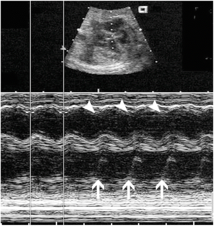
Fig. 20.7
M-mode echocardiogram from a fetus with supraventricular tachycardia. Arrowheads indicate atrial contractions, and large arrows indicate ventricular contractions. Note the 1:1 relationship of atrial and ventricular contractions. The calipers measure 243 ms between successive ventricular contractions, indicating a heart rate of approximately 250 bpm
Treatment is predicated on the effect of the tachyarrhythmia on fetal well-being. If SVT is intermittent and late in pregnancy, fetal health is usually not jeopardized. In such patients, prognosis is generally excellent, and no treatment is indicated. If tachycardia is sustained at fast rates (>260 bpm), prenatal morbidity and even demise may be as high as 25 %. It is therefore important to frequently (every 3–5 days) assess all fetal patients who remain in SVT. Both obstetricians and cardiologists should be involved in the care of patients. Of note, patients with structural congenital heart disease are at greater risk for tachycardia-associated complications. One of the earliest signs of fetal compromise is an exaggerated systemic venous (inferior vena cava or hepatic) flow reversal (greater than 30 %) on the venous Doppler. This may progress to reversal in the ductus venosus, and eventual notching in the umbilical venous tracing in more advanced fetal heart failure. Other signs of fetal distress include cardiomegaly, decreased ventricular systolic function, atrioventricular valve regurgitation, and hydrops fetalis (pericardial effusion, pleural effusion, ascites, and/or skin edema).
Fetal treatment options include early delivery, transplacental (maternal) pharmacotherapy, or direct fetal pharmacotherapy. In addition, there is one report of fetal SVT conversion using trans-abdominal umbilical cord compression. Although not described in humans, there has been a single report of fetal SVT conversion using fetal transesophageal pacing in fetal sheep. Labor induction with delivery is the treatment of choice for term and near-term pregnancies with sustained fetal tachycardia or evidence of fetal compromise.
Although controversy exists, it is generally agreed upon that transplacental digoxin should be the first-line treatment of choice in pre-term pregnancies with sustained tachycardia without fetal compromise (Table 20.1). Digoxin treatment is safe and often effective. The drug can be administered to the mother in oral or IV form at relatively high maternal doses (up to 1 mg q day orally) in order to achieve an adequate level in the fetus. SVT cessation is achieved in approximately three-quarters of cases with maternal oral therapy. If conversion has not been achieved after 2 weeks of therapy with adequate maternal digoxin levels (1–2 ng/mL), a second antiarrhythmic agent may be added. Flecainide, along with other drug choices are reasonable (Table 20.1). When there are signs of fetal compromise, it is reasonable to begin dual therapy immediately (digoxin along with a second-line agent such as flecainide). Several studies in the literature as well as clinical experience have suggested that transplacental transfer of digoxin is limited in the hydropic fetus, whereas placental transfer of flecainide and sotalol are not as affected. Cardioversion with flecainide is generally achieved within 3–4 days. Other medications with reported efficacy include procainamide, verapamil, quinidine, amiodarone, or sotalol. There have been no definitive large, randomized published studies comparing fetal antiarrhythmic agents. Success with sotalol or the combination of digoxin with amiodarone has been described.
Table 20.1
Pharmacotherapy for treatment of fetal tachyarrhythmia
Drug | Route of administration | Starting dosage | Maximum dosage | SVT | AFL | VT | Maternal/fetal adverse effects |
|---|---|---|---|---|---|---|---|
Adenosine | Fetal IV | 0.1 mg/kg | 0.2 mg/kg | Yes | No | No | Tachycardia commonly recurs requiring additional agent |
Amiodarone | Maternal PO (fetal IV; fetal IM; fetal IP) | 800–1,200 mg QD X 2–7 days load; then 200–800 mg QD maint. | 800 mg QD maint. | Yes | No | Yes | Maternal or fetal hypothyroidism (common); bradycardia (common); IUGR; premature delivery; hepatotoxicity; monitor maternal QTc |
Digoxin | Maternal PO; maternal IV (fetal IV; fetal IM; fetal IP) | PO: 0.25–1.5 mg load, then 0.125–0.25 mg PO BID-TID maint. < div class='tao-gold-member'>
Only gold members can continue reading. Log In or Register to continue
Stay updated, free articles. Join our Telegram channel
Full access? Get Clinical Tree
 Get Clinical Tree app for offline access

|