Segmental Anatomy

Chapter 106 Segmental Anatomy


SEGMENTAL SETS OR COMBINATIONS


The cardiac segments are the anatomic and developmental building blocks out of which all human hearts, normal and abnormal, are made.18 The three main cardiac segments are the atria, the ventricles, and the great arteries (Fig. 106-1). The two connecting cardiac segments are the atrioventricular canal or junction, and the infundibulum, or conus arteriosus (see Fig. 106-8). Understanding segmental anatomy1 requires an understanding of morphologic anatomy3: the anatomic features of the morphologically right atrium (RA) (Fig. 106-2), of the morphologically left atrium (LA) (Fig. 106-3), of the morphologically right ventricle (RV) (Fig. 106-4), and of the morphologically left ventricle (LV) (Fig. 106-5). As Figure 106-1 illustrates, the morphologically RA, LA, RV, and LV can positionally be anywhere. A combination of morphologic anatomy and segmental anatomy is the basis of accurate diagnosis in congenital heart disease (see Fig. 106-1), which in turn is basic to successful open-heart surgery.


image

Figure 106–1 Types of human heart in terms of segmental sets (combinations) and segmental alignments. Heart diagrams are viewed from below, as would be achieved with a subxiphoid two-dimensional echocardiogram. Cardiotypes depicted in broken lines had not been documented when this diagram was made. The aortic valve is indicated by the coronary ostia. The pulmonary valve is indicated by the absence of coronary ostia. Braces {} mean “the set of.” The columns (1 to 4) are arranged in terms of atrioventricular (AV) concordance or discordance. Column 1 (i.e., {S,D,-}) has AV concordance in visceroatrial situs solitus. Column 2 (i.e., {S,L,-}) has AV discordance in visceroatrial situs solitus. Column 3 (i.e., {I,L,-}) has AV concordance in visceroatrial situs inversus. Column 4 (i.e., {I,D,-}) has AV discordance in visceroatrial situs inversus. Situs ambiguus of the viscera and atria in the heterotaxy syndromes, particularly with asplenia (i.e., {A,D,-} and {A,L,-}) is omitted; the concepts of AV concordance and AV discordance do not apply in visceroatrial situs ambiguus because the frame of reference, the type of visceroatrial situs, is uncertain or unknown. The rows (1 to 8) are organized in terms of the types of ventriculoarterial (VA) alignment. Normal concordant VA alignments are depicted in rows 1 to 4. Abnormal concordant VA alignments are shown in row 6 and concern anatomically corrected malposition of the great arteries. Discordant VA alignments are depicted in row 5 and concern transposition of the great arteries. Double outlets are shown in rows 7 and 8, double-outlet right ventricle in row 7, and double-outlet left ventricle in row 8. All associated malformations are omitted for diagrammatic simplicity and clarity. Ant, anterior; Inf, infundibulum; LA, morphologically left atrium; L, left; LV, morphologically left ventricle; Post, posterior; R, right; RA, morphologically right atrium; RV, morphologically right ventricle.


(From Foran RB, Belcourt C, Nanton MA, et al. Isolated infundibuloarterial inversion {S,D,I}: a newly recognized form of congenital heart disease. Am Heart J 1988;116:1337-50.)


image

Figure 106–2 Morphologically right atrium (RA). A, Exterior. B, Interior. In A, note the large, broad, triangular right atrial appendage (RAA). The RA always receives the inferior vena cava (IVC) and normally receives the superior vena cava (SVC). Note the groove or sulcus where the SVC enters the RA; this is the sulcus terminalis (terminal sulcus), where the sinoatrial node (SA; the pacemaker of the heart) is located. In B, note the IVC, which is a highly reliable diagnostic marker of the RA, as is the ostium of the coronary sinus (CoS). The SVC is not a highly reliable diagnostic marker of the RA because the SVC may open into the morphologically left atrium (LA) when the coronary sinus is unroofed, resulting in a coronary sinus septal defect. The septal surface of the RA is characterized by the superior limbic band of septum secundum (Sept II) (Latin septum secundum, “the second septum”). Septum primum (Sept I) is the flap valve of the foramen ovale (oval foramen) that normally opens into the LA prenatally (Latin septum primum, “the first septum”). The crista terminalis (CT) lies lateral to the entry of the SVC (Latin crista terminalis, “terminal crest”). The CT corresponds internally to the sulcus terminalis externally where the SA node and the SA nodal artery are located. Deep sutures into the CT should be avoided so as not to damage the SA node and the SA nodal artery, causing sick sinus syndrome. The musculi pectinati (MP) are prominent over the interior of the RAA. Musculi pectinati means pectinate muscles, these parallel muscular ridges resembling the teeth of a comb (Latin pectin, “comb”). The tinea sagittalis (TS) is also seen (Latin tinea sagittalis, “sagittal worm”). Catheter tips can get lodged between the TS and the CT, where an injudicious push of the catheter can lead to perforation of the RA and cardiac tamponade. The anterior (Ant), posterior (Post), and septal (Sept) leaflets of the tricuspid valve (TV) are seen from above. The membranous septum is located at the commissure between the Ant and Sept leaflets of the TV. The atrioventricular (AV) node and the bundle of His of the AV conduction system are, unfortunately, invisible; consequently, the cardiac surgeon must know where they are. Draw a mental line—not with a sucker that can get attached to the endocardium and when pulled away can cause heart block—between the ostium of the CoS and the membranous septum (MS) at the Ant leaflet–Sept leaflet commissure. Beneath the MS is where the proximal unbranched portion of the AV bundle dives, becoming the penetrating portion of the AV bundle that then passes anteroinferiorly to reach the ventricles, where it divides into the left and right bundle branches. The coronary sinus–to–membranous septum line is exactly where the AV node and the proximal unbranched portion of the AV bundle are located. The CoS-MS line is particularly helpful surgically when the valves that are supposed to delimit the triangle of Koch, in which the AV node and unbranched AV bundle are located, happen to be absent, or are difficult to see, or are abnormally located: the eustachian valve of the IVC and its anterior invisible extension, the tendon of Todaro; the thebesian valve of the CoS; and the origin of the Sept leaflet of the TV. Even when these valve leaflets outlining the triangle of Koch are unhelpful, the CoS-MS line can be used to localize the invisible AV node and the unbranched portion of the AV bundle. Ao, ascending aorta; PA, main pulmonary artery; RV, exterior of the morphologically right ventricle.


Rights were not granted to include this content in electronic media. Please refer to the printed book.


(From Van Praagh R, Vlad P. Dextrocardia, mesocardia, and levocardia: the segmental approach to diagnosis in congenital heart disease. In: Keith JD, Rowe RD, Vlad P, editors. Heart disease in infancy and childhood. 3rd ed. New York: Macmillan; 1978, p. 638-95.)


image

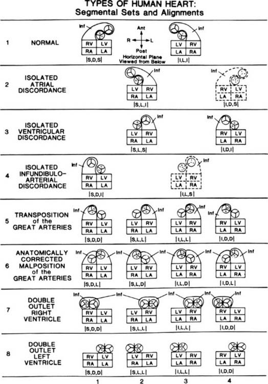
Figure 106–3 Morphologically left atrium (LA). A, Exterior. B, Interior. In A, the left atrial appendage (LAA) is relatively long and thin, like a pointing finger. It is also small and posterior, compared with the right atrial appendage (RAA). Normally, the LAA is not incorporated into the main cavity of the LA, whereas the RAA is incorporated to a major degree into the main cavity of the RA. The pulmonary veins (PV) normally connect with the LA; however, abnormally, the PV do not return to the LA, as in totally anomalous pulmonary venous connection. Hence, the pulmonary veins are not a highly reliable diagnostic marker of the LA. To our knowledge, the IVC never connects directly with the LA. Rarely, the IVC can connect with the left sinus horn (an enlarged coronary sinus [CoS]), and if a coronary sinus septal defect coexists, unroofing the CoS, then the bloodstream of the IVC can flow into the LA. However, note that the IVC connects with the left sinus horn, not with the LA directly. In B, note that septum primum (Sept I) normally is the dominant morphologic feature of the LA’s septal surface. Septum secundum (Sept II) is partly covered by the more leftward Sept I. The pectinate muscles of the LA are largely confined to the small fingerlike LAA and do not normally extend out into the main cavity of the LA. Consequently, the pectinate muscles of the LA do not approach the mitral valve (MV), whereas the pectinate muscles of the RA closely approach the tricuspid valve (see Fig. 106-2B). This characteristic difference in distribution of papillary muscles in the LA compared with the RA reflects the different degrees of incorporation of the atrial appendages and their pectinate muscles into the main chamber: in the LA, pectinate incorporation is minimal; in the RA, pectinate incorporation is maximal. Differing degrees of incorporation of the atrial appendages are reflected by differing pectinate patterns; this understanding is important in the heterotaxy syndromes with asplenia and polysplenia.


Rights were not granted to include this content in electronic media. Please refer to the printed book.


(From Van Praagh R, Vlad P. Dextrocardia, mesocardia, and levocardia: the segmental approach to diagnosis in congenital heart disease. In: Keith JD, Rowe RD, Vlad P, editors. Heart disease in infancy and childhood. 3rd ed. New York: Macmillan; 1978, p. 638-95.)


image

Figure 106–4 Morphologically right ventricle (RV). A, Exterior, drawing. B, Interior, drawing. C, Interior, photograph. In A, note that the exterior of the RV is triangular, as is the exterior of the right atrium (RA). The anterior and diaphragmatic surfaces of the RV form an acute angle with one another of less than 90 degrees; hence, this is called the acute margin of the heart, and the branch of the right coronary artery that runs along it is called the acute marginal (M) branch. Two or more preventricular branches of the right coronary artery are also typical of the RV. The anterior descending (AD) branch of the left coronary artery indicates the location of the ventricular septum, to the left of which lies the morphologically left ventricle (LV). The main pulmonary artery (PA) normally arises anteriorly, superiorly, and to the left of the ascending aorta (Ao), which originates posteriorly, inferiorly, and to the right of the PA. In B and C of the interior of the RV, note that the conal septum (CS) and its parietal or free-wall extension, the parietal band (PB), separate the normally located pulmonary valve above from the tricuspid valve (TV) below. The septal band (SB) lies on the right ventricular septal surface anterosuperiorly. The SB gives off the moderator band (MB), which carries the right bundle branch of the AV conduction system from the septum medially to the anterior papillary (AP) muscle of the RV laterally. From there, the right bundle branch arborizes on the interior of the RV free wall as the Purkinje network. The muscle of Lancisi (ML), also known as the muscle of Luschka and as the medial papillary muscle, anchors the anterior and the septal leaflets of the tricuspid valve (TV) anterosuperiorly. This is the anterosuperior commissure of the TV, where the membranous septum (pars membranacea septi) is located. The membranous septum lies immediately behind or medial to the ML. Just below the ML is where the right bundle branch (RBB) of the conduction system emerges into the RV.


The RBB then runs down the SB, close to its inferior and posterior margin, heading toward the MB. Often it is possible to see the RBB with the naked eye, without special stains, as a thin, yellow-white line. Surgically, this means that it is often possible to avoid transfixing the RBB with surgical needles or sutures during ventricular septal defect patch placement. Thus it is often possible to avoid real RBB block—as opposed to arborization block, which resembles true RBB block in the surface electrocardiogram but not in the intracardiac electrocardiogram. The TV attaches to the RV septal surface via multiple chordae tendineae (tendinous chords). Hence, the TV is “septophilic.” By contrast, the mitral valve is “septophobic”—an important diagnostic point that distinguishes the RV from the LV (in prenatal echocardiograms, e.g.). The trabeculae carneae (fleshy ridges) of the RV are relatively few, coarse, and straight, very different from those of the LV, which are numerous, fine, and oblique. The RV sinus, body, or inflow tract—the main pumping portion—lies beneath or behind (depending on whether the heart position is horizontal, as in infants and children, or vertical, as in many adults) the infundibulum, conus, or outflow tract. A ring of conal or infundibular muscle formed by the CS, PB, SB, and MB demarcates the RV sinus (inflow tract) upstream, from the conus (outflow tract) downstream. The main pumping portion of the RV—the inflow tract—is relatively small. The outflow tract or conus is not a good pump. The main function of the normal conus is structural, to cross the pulmonary and systemic circulations during normal cardiogenesis. The conus normally performs this embryonic arterial switch operation. The ventricles can usually be identified by external inspection on the basis of their distinctive shapes and coronary arterial patterns. The CS is also known as the outlet septum. The PB is also called the ventriculo-infundibular fold. The SB and MB are also called the trabecula septomarginalis or septomarginal trabeculation.


Rights were not granted to include this content in electronic media. Please refer to the printed book.


(From Van Praagh R, Vlad P. Dextrocardia, mesocardia, and levocardia: the segmental approach to diagnosis in congenital heart disease. In: Keith JD, Rowe RD, Vlad P, editors. Heart disease in infancy and childhood. 3rd ed. New York: Macmillan; 1978, p. 638-95.)


image

Figure 106–5 Morphologically left ventricle (LV). A, Exterior, drawing. B, Interior drawing. C, Interior, photograph. In A, note that the external shape of the LV is conical, like a bullet or a torpedo. The lateral margin of the LV is described as the obtuse margin because it forms an angle of more than 90 degrees (as opposed to the acute margin of the right ventricle, Fig. 106-4A). The anterior descending (AD) coronary artery and the posterior descending coronary artery (not seen) externally indicate the location of the ventricular septum. The diagonal (D) branches of the left coronary artery, also known as the obtuse marginal branches, externally indicate the locations of the left ventricular papillary muscles. The superior diagonal supplies the region of the anterolateral (AL) papillary muscle group and the adjacent LV free wall. The inferior diagonal supplies the region of the posteromedial papillary (PP) muscle group and the adjacent LV free wall. Also seen are the characteristic external shape of the left atrium (LA), the leftward and anterior location of the normally arising main pulmonary artery (PA), and the more rightward and posteroinferior location of the normally originating ascending aorta (Ao). In B and C, the noncoronary (NC) and left coronary (LC) leaflets of the aortic valve are in direct fibrous continuity with the anterior leaflet of the mitral valve (AL of MV), reflecting the normal absence of the subaortic conal free-wall myocardium. The anterosuperior portion of the left ventricular septal (S) surface is smooth or nontrabeculated. The inferior and apical portions of the left ventricular septal and free-wall (FW) surfaces are characterized by a lattice-like mesh of small, fine, oblique trabeculae carneae (very different from the coarse, few, straight trabeculations of the RV). The ostia of the right coronary (RC) and LC arteries are seen in their respective sinuses of Valsalva. The noncoronary–left coronary commissure normally is located above the middle of the anterior mitral leaflet. The noncoronary–right coronary commissure normally is located above the membranous septum, which in turn is immediately above the left bundle of the atrioventricular (AV) conduction system.


An understanding of these normal landmarks can be very helpful surgically—for example, when doing a transaortic valvar resection of discrete fibrous subaortic stenosis. The commissures of a normally related aortic valve help to indicate to the surgeon where these various important, initially invisible, and vulnerable subaortic structures are located. Often, one can see the superior (anterior) and the inferior (posterior) radiations of the left bundle branch of the AV conduction system with the naked eye, without special stains, as shown in B. Being glycogen rich, these yellow-white superior and inferior radiations often can be seen arching across the smooth left ventricular septal surface, the superior radiation running to the superior (AL) papillary muscle group and the inferior radiation running to the inferior (PP) papillary muscle group. The left ventricular papillary muscles are few, large, and they arise from the left ventricular free wall—never normally from the left ventricular septal surface. Hence, the papillary muscles of the LV are “septophobic”—the opposite of those of the RV, which are “septophilic.”


Rights were not granted to include this content in electronic media. Please refer to the printed book.


(From Van Praagh R, Vlad P. Dextrocardia, mesocardia, and levocardia: the segmental approach to diagnosis in congenital heart disease. In: Keith JD, Rowe RD, Vlad P, editors. Heart disease in infancy and childhood.3rd ed. New York: Macmillan; 1978, p. 638-95.)


For each type of heart (see Fig. 106-1), the atria, ventricles, and great arteries may be regarded as the members of a set or combination: {atria, ventricles, great arteries}. Braces {} are a mathematical symbol meaning “the set of.” The main cardiac segments are the members of a segmental set or combination. Each segmental set is recorded in venoarterial sequence, {atria, ventricles, great arteries}, this being the natural or blood-flow order.


MORPHOLOGIC ANATOMY


Beginning with the atria, two types of visceroatrial situs are shown in Figure 106-1.9 Situs means the pattern of anatomic organization. We speak of visceroatrial situs because the situs of the viscera and the situs of the atria are almost always the same. The two types of visceroatrial situs (see Fig. 106-1) are (1) situs solitus, the usual, ordinary, customary situs—hence the normal; and (2) situs inversus, a mirror-image of situs solitus. In a mirror image, there is right–left reversal without anteroposterior or superoinferior change. Visceroatrial situs solitus and situs inversus are stereoisomers.


A third possibility is situs ambiguus,3,1012 the uncertain or indeterminate type of visceroatrial situs that may characterize the heterotaxy syndromes with asplenia, with polysplenia, or with a normally formed but often right-sided spleen. Visceroatrial situs ambiguus, which can occur with these anomalies of laterality—the heterotaxy syndromes,12 is not regarded as a specific type of visceroatrial situs. Instead, situs ambiguus appears to be a scrambled form either of visceroatrial situs solitus or of visceroatrial situs inversus. As noted earlier, this is why there appear to be only two types of visceroatrial situs: solitus and inversus (see Fig. 106-1).


It should be understood that the concepts of atrial isomerism and atrial appendage isomerism are considered to be erroneous.12,13 To be accurate, atrial isomerism has never been documented—neither bilateral RA, with bilateral inferior venae cavae, bilateral superior venae cavae, bilateral coronary sinus ostia, bilateral septa secunda, and bilateral broad triangular appendages; nor bilateral LA with bilateral pulmonary veins, bilateral septa prima, and bilateral fingerlike appendages.


Similarly, isomerism of parts of the atria, such as isomerism of the pectinate muscles (only), is regarded as a contradiction in terms.13 Isomerism applies to a whole structure, not just to a few of its parts. For example, the molecules D-glucose and L-glucose would not be regarded as isomers if only some of the hydroxyl groups, or some of the hydrogen atoms, or only the carboxyl groups were mirror images but nothing else was. Thus, pectinate isomerism is considered to be an erroneous concept because isomerism applies to whole structures, such as the RA and the LA, not just to parts of a structure, such as the pectinate muscles.


The realization that atrial-level isomerism is an erroneous concept facilitates the diagnosis of atrial situs, even in the heterotaxy syndromes.12 However, when atrial situs cannot be diagnosed with confidence, we make the diagnosis of atrial situs ambiguus, meaning that the atrial situs is undiagnosed, but without the implication that atrial-level isomerism is present.


In visceroatrial situs solitus, symbolized as {S,-,-} (see Fig. 106-1, columns 1 and 2), the RA (see Fig. 106-2) is right-sided and the LA (see Fig. 106-3) is left-sided. In visceroatrial situs inversus, symbolized as {I,-,-} (see Fig. 106-1, columns 3 and 4), the mirror-image RA is left-sided and the mirror-image LA is right-sided.


There are two types of ventricular situs (isomeric patterns of anatomic organization): D-loop ventricles (see Fig. 106-1, columns 1 and 4) and L-loop ventricles (see Fig. 106-1, columns 2 and 3). D-loop ventricles are solitus or noninverted ventricles that are normally associated with dextral or rightward looping of the straight heart tube, placing the RV (see Fig. 106-4) to the right of the LV (see Fig. 106-5). L-loop ventricles are inverted or mirror-image ventricles that are typically associated with levo or leftward looping of the straight heart tube, placing the mirror-image RV to the left of the mirror-image LV.


CHIRALITY OR HANDEDNESS


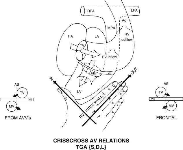
The D-loop, or solitus RV, is right-handed (Fig. 106-6).14 Figuratively speaking, the thumb of the right hand goes through the tricuspid valve. The fingers of the right hand go into the RV outflow tract. The palm of only the right hand faces the RV septal surface. The dorsum of only the right hand faces the RV free-wall surface.


image

Figure 106–6 The D-loop right ventricle (RV) is right-handed. Figuratively speaking, the thumb of the right hand goes through the tricuspid valve (TV) and indicates the location of the RV inflow tract (IN). The fingers of the right hand go into the RV outflow tract (OUT). The palm of only the right hand faces the RV septal surface, and the dorsum of only the right hand is adjacent to the RV free-wall surface. Because the D-loop RV is right-handed, D-loop ventricles are also known as right-hand topology. Handedness or chirality is helpful in making the diagnosis of the ventricular situs (i.e., D-loop or solitus ventricles versus L-loop or inversus ventricles) when the conventional definitions of noninversion and inversion relative to the sagittal plane break down, as in this case. The patient illustrated by this diagram had superoinferior ventricles with a superior RV, an inferior morphologically left ventricle (LV), and an approximately horizontal ventricular septum (VS). Crisscross atrioventricular (AV) relations were also present; note the approximately 90-degree angle between the right atrial (RA)–to–RV inflow tract (solid white arrow) and the left atrial (LA)–to–LV inflow tract (broken white arrow). Both ventricles are bilateral, right-sided and left-sided, which is why the conventional definition of noninversion versus inversion does not work. The RV begins superiorly on the right beneath the TV, and then it proceeds leftward and is mostly left-sided. The LV begins inferiorly on the left beneath the mitral valve (MV) and then proceeds rightward and is mostly right-sided. Note the view of the atrial septum (AS), the VS, the TV, the MV, and the opposite directions of the AV inflow tracts (black arrows) as seen from the AV valves (from AVV’s), which is the surgeon’s perspective, or from the front (FRONTAL), which is the echocardiographer’s view in the short-axis projection. Crisscross heart results from wrong-way ventricular looping or twisting. After concordant AV alignments and connections have been established, these D-loop ventricles twisted leftward—instead of rightward, which is normal for D-loop ventricles. Hence, the atria and the ventricles are mostly contralateral (instead of being ipsilateral, which is normal): the RA is right-sided, but the RV is mostly left-sided. The LA is left-sided, but the LV is mostly right-sided. Hence, there was an appearance of atrioventricular contralaterality (instead of the usual atrioventricular ipsilaterality). The diagnosis in this patient was transposition of the great arteries (TGA) with the segmental set of {S,D,L}, meaning situs solitus of viscera and atria {S,-,-}, with D-loop ventricles {S,D,-}, and L-TGA (aortic valve levo, or L, relative to the pulmonary valve) (i.e., {S,D,L}). There was AV concordance with ventriculoarterial (VA) discordance. Because there was one segmental discordance (at the VA level), the systemic and pulmonary venous circulations were physiologically uncorrected. Both venous circulations had physiologically inappropriate destinations: systemic venous blood to the aorta (Ao), and pulmonary venous blood to the main pulmonary artery (MPA). Hence, TGA {S,D,L}was “complete,” or physiologically uncorrected. The transposed aortic valve was anterior and to the left of the transposed pulmonary valve because there was a well-developed subaortic conus, with pulmonary-mitral fibrous continuity, indicating resorption of the subpulmonary conal free wall. With superoinferior ventricles and a horizontal ventricular septum, the ventricles were “lying down.” If one “corrects” this ventricular malposition by making the ventricles “sit up” approximately normally (i.e., with a vertical VS instead of a horizontal VS), then the transposed aortic valve becomes superior and rightward relative to the transposed pulmonary valve. Hence, the leftward location of the transposed aortic valve in TGA {S,D,L} is caused by the subaortic conus plus the abnormally horizontal ventricular malposition. This ventricular malposition also causes the horizontal VS, the superoinferior ventricles, and the crisscross AV relationships. Note that even when the ventricles are very malpositioned, as in this patient, when the TV is right-sided (and superior) and when the MV is left-sided (and inferior), D-loop ventricles are present. The right–left relationships of the TV and the MV are very highly reliable indicators of which kind of ventricular loop is present: right-sided TV indicates D-loop ventricles, and left-sided TV indicates L-loop ventricles. LPA, left pulmonary artery; RPA, right pulmonary artery.


(From Van Praagh S, LaCorte M, Fellows KE, et al. Superoinferior ventricles, anatomic and angiocardiographic findings in 10 postmortem cases. In: Van Praagh R, Takao A, editors. Etiology and morphogenesis of congenital heart disease. Mt. Kisco, NY: Futura: 1980, p.  317-78.)

< div class='tao-gold-member'>

Stay updated, free articles. Join our Telegram channel

Jul 30, 2016 | Posted by in CARDIAC SURGERY | Comments Off on Segmental Anatomy

Full access? Get Clinical Tree

Get Clinical Tree app for offline access