Respiratory Syncytial Virus Infection (Bronchiolitis or Pneumonitis)


Respiratory Syncytial Virus Infection (Bronchiolitis or Pneumonitis)


Anatomic Alterations of the Lungs


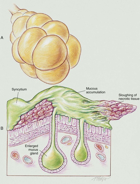
The respiratory syncytial virus (RSV) moves down the respiratory tract by means of cell-to-cell transfer, causing bronchiolitis and, later, atelectasis and pneumonia in the child. The syncytium is defined as a “multinucleate mass of protoplasm produced by the merging of cells.” At the level of the bronchioles the virus causes neighboring cells to fuse to form a syncytium, hence the name respiratory syncytial virus. The lower airways may also become infected when secretions from RSV-infected upper airways are aspirated.


RSV infection causes peribronchiolar mononuclear infiltration and necrosis of the epithelium of the small airways. This condition leads to edema of the small airways and increased production of mucus. As the condition worsens, the epithelium of the small airways becomes necrotic and proliferates into the airway lumen. The combination of sloughing necrotic tissue, airway edema, and accumulation of mucus leads to (1) a decreased airway lumen, (2) a partially obstructed airway, or (3) a completely obstructed airway. Partial airway obstruction leads to alveolar hyperinflation as a result of a “ball-valve” mechanism (see Figure 36-1). Complete airway obstruction leads to alveolar collapse or atelectasis. Pneumonic consolidation is common. RSV is also referred to as bronchiolitis or pneumonitis.


The following major pathologic or structural changes are associated with RSV infection:


Etiology and Epidemiology


RSV is the most common viral respiratory pathogen seen in infancy and early childhood. Although RSV infection can occur at any age, it is most commonly seen in young children. Almost all children will be infected with RSV by their second birthday. At highest risk for severe respiratory distress syndrome (RDS) are premature infants, children less than 1 year of age, and children with weakened immune systems as a result of a medical condition or medical treatment. Adults with compromised immune systems and those 65 years of age and older are also at increased risk of severe RDS.


RSV is commonly transmitted by young children who are infected with RSV and demonstrate the signs and symptoms of a mild upper respiratory tract infection or a cold—for example, coughing, sneezing, runny nose, decreased appetite, irritability, decreased activity, and respiratory distress. RSV is easily transmitted when droplets containing the virus are coughed or sneezed into the air. Infection occurs when the particles touch the nose, mouth, or eyes of uninfected individuals in the immediate area. RSV can also spread from direct or indirect contact with nasal or oral secretions from an infected person. For example, RSV can be contracted by kissing the face or hands of a child infected with RSV who has a runny nose. Indirect contact may occur when touching the hard surface of a table, crib rail, or doorknob that has been touched by a person infected with RSV. RSV can survive several hours on a hard surface. Common areas of RSV transmission include elementary schools and day care centers. Frequent hand washing and wiping the hard surfaces with a disinfectant may help stop the spread of RSV.


Infants and children infected with RSV usually develop symptoms within 4 to 6 days of infection (range: 2 to 8 days). Most will recover in 1 to 2 weeks. Infected individuals are usually contagious for 3 to 8 days. However, even after recovery an infected person can spread the virus for 1 to 3 weeks. Some patients with a weakened immune system may be contagious for as long as 4 weeks.


Most otherwise healthy children with RSV do not require hospitalization. However, according to the Centers for Disease Control and Prevention, 75,000 to 125,000 children under the age of 1 year are hospitalized each year in the United States because of RSV infection. Of this group, most of the children hospitalized because of RSV are under 6 months old.


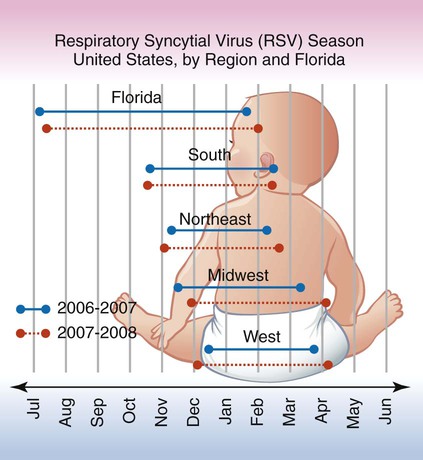
Although the outbreak of RSV cases varies by location each year, the number of RSV cases typically increases during the fall, winter, and early spring months. It is not fully known why RSV outbreaks occur in certain regions more than in others, but temperature, climate, and humidity appear to play a role. Figure 36-2 shows the typical RSV season in the United States by region and in Florida according to the Centers for Disease Control and Prevention.


Laboratory Testing for Respiratory Syncytial Virus


RSV infection should be suspected when the clinical manifestations correlate to the time of year, the presence of a local outbreak, the age of the patient, and the history of the illness. Through use of an oropharyngeal or nasopharyngeal secretion sample, RSV is most commonly diagnosed with commercially available antigen assay tests. RSV can also be confirmed with a nasopharyngeal culture. Both the antigen assay test and the nasopharyngeal culture are usually reliable in young children but are less sensitive in older children and adults. Highly sensitive reverse transcriptase–polymerase chain reaction (RT-PCR) assays are also available. The RT-PCR assay may be used to test adults.


Jun 11, 2016 | Posted by in RESPIRATORY | Comments Off on Respiratory Syncytial Virus Infection (Bronchiolitis or Pneumonitis)

Full access? Get Clinical Tree

Get Clinical Tree app for offline access