Epicardial adipose tissue (EAT) contains abundant ganglionated plexi that might contribute to the occurrence of atrial fibrillation (AF). Maximal standardized uptake value (SUV) of 18-fluorodeoxyglucose (FDG)-positron emission tomography (PET) reflects glucose metabolism of the tissue. It has also been shown that FDG-PET is proportional to macrophage density. We examined EAT inflammatory activity using FDG-PET/computerized tomography in patients with AF and in controls. Retrospective analysis of patients who underwent FDG-PET/computerized tomography was performed. About 21 consecutive patients with confirmed history of AF and 21 non-AF control group matched for age, gender, and body mass index (BMI) were included. SUV was measured in fat adjacent to the roof of left atrium, right ventricle, atrioventricular groove, and left main artery. Additionally SUV was measured in subcutaneous fat and visceral thoracic fat. In both groups, associations of SUV with gender, age, BMI, and serum glucose were further analyzed. EAT SUV measured near the roof of left atrium, atrioventricular groove, and left main artery was significantly greater in patients with AF than in control group (1.66 ± 0.36 vs 1.23 ± 0.32, p = 0.00015; 2.07 ± 0.50 vs 1.51 ± 0.24, p = 0.00003; and 1.95 ± 0.48 vs 1.52 ± 0.26, p = 0.0007, respectively). In addition, EAT SUV was significantly greater than subcutaneous and visceral thoracic fat for patients with AF and controls. EAT SUV was not related to gender, age, BMI, or serum glucose. In conclusion, inflammatory activity of EAT reflected by SUV is higher in patients with AF than that in controls. Inflammatory activity of EAT adjacent to left atrium, atrioventricular groove, and left main artery is greater than in subcutaneous or visceral thoracic tissue.
The aim of our study was to compare epicardial adipose tissue (EAT) inflammatory activity using 18-fluorodeoxyglucose-positron emission tomography/computerized tomography in patients with atrial fibrillation (AF) and in controls. We also wanted to test relation of EAT inflammatory activity with gender, age, body mass index (BMI), and serum glucose level.
Methods
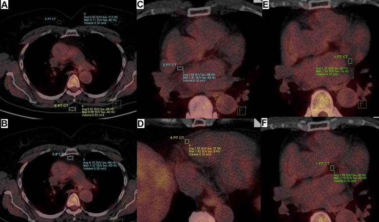
Retrospective case–control analysis was performed. Out of patients who underwent fluorodeoxyglucose-positron emission tomography/computerized tomography due to suspicion of neoplastic disease in the years 2008–2010, 21 consecutive patients with confirmed history of AF (paroxysmal = 9 and persistent = 12) and 21 patients without history of AF, matched for age, sex, and BMI (control group), were selected. Standardized uptake value (SUV) was measured in fat adjacent to the roof of left atrium, right ventricle, atrioventricular groove, and left main coronary artery ( Figure 1 ). Additionally SUV was measured in subcutaneous fat and intrathoracic (visceral) fat—nonepicardial fat. Associations of SUV with gender, age, BMI, and serum glucose were further analyzed. Mean of 3 measurements was taken by 2 researchers blind to result with interobserver and intraobserver variabilities of <5%.

Normality of distribution was tested with Shapiro-Wilk test. Continuous variables are presented as mean ± SD, and categorical variables are presented as frequencies. Analysis of variance was used to test differences between SUV in different locations. Differences between AF and control groups are calculated with student t or Chi-square and Fisher’s exact tests, according to variable and its distribution. Seven different measurements of adipose tissue inflammatory activity were measured, so after Bonferroni correction, p value <0.007 (0.05/7) was considered statistically significant in univariate analysis. In multivariate analysis, several parameters were included: age, gender, BMI, hypertension, and SUV in different locations; p <0.05 was considered statistically significant. All analyses were performed with Statistica 10 (Statsoft, Inc., Tulsa, Oklahoma).
Results
Characteristic of the total population with the division on AF and control groups is listed in Table 1 . AF and control groups were not different with respect to most of the clinical and demographic parameters. SUV measured in fat adjacent to the roof of left atrium, right ventricle, atrioventricular groove, and left main coronary artery was significantly greater than SUV in subcutaneous and visceral fat for patients with AF and healthy controls (p <0.01).
| Variable | Total (n = 42) | AF Group (n = 21) | Control Group (n = 21) | p Value |
|---|---|---|---|---|
| Age (yrs) | 72.2 ± 7.2 | 73.3 ± 7.4 | 71.0 ± 7.0 | 0.31 |
| Male gender | 21 (50) | 11 (52) | 10 (48) | 1.00 |
| Body mass index (kg/m 2 ) | 27 ± 5 | 28 ± 4 | 27 ± 6 | 0.66 |
| Body surface area (m 2 ) | 2.77 ± 0.3 | 2.75 ± 0.33 | 2.79 ± 0.33 | 0.69 |
| Hypertension | 26 (62) | 14 (67) | 12 (57) | 0.75 |
| Diabetes mellitus | 7 (17) | 4 (19) | 3 (14) | 0.70 |
| Coronary artery disease | 13 (31) | 7 (33) | 6 (28) | 1.00 |
| Previous myocardial infarction | 8 (19) | 4 (19) | 4 (19) | 1.00 |
| Congestive heart failure | 11 (26) | 6 (29) | 5 (24) | 1.00 |
| Neoplastic disease ∗ | 24 (55) | 11 (52) | 13 (62) | 0.76 |
| β blockers | 31 (74) | 16 (76) | 15 (71) | 1.00 |
| ACEI/ARB | 27 (64) | 15 (71) | 12 (57) | 0.51 |
| Aspirin | 13 (31) | 4 (19) | 9 (43) | 0.18 |
| Statins | 21 (50) | 10 (48) | 11 (52) | 1.00 |
∗ Suspicion of colon or prostate cancer (not confirmed); no other known inflammatory conditions were present; no other anti-inflammatory medicines were taken during examination.
Major results of the study are listed in Table 2 . EAT SUV measured in fat adjacent to the roof of left atrium, atrioventricular groove, and left main coronary artery was substantially greater in patients with AF than in control group, and the difference was statistically highly significant. SUV measured in other locations was not different between groups. Paroxysmal versus persistent AF, duration of AF, left atrium size, as well as serum C-reactive protein levels did not correlate with EAT SUV. EAT SUV was not related to gender, age, BMI, body surface area, or serum glucose (data not shown). In multivariate logistic regression analysis, we included SUV near the roof of left atrium (representing all pericardial tissue—due to significant correlation between variables), SUV in subcutaneous posterior, and SUV in visceral fat. Only SUV near the roof of left atrium (odds ratio 116.80, 95% confidence interval 5.70 to 2,394.14, p = 0.002) was significant predictor of AF in multivariate logistic regression analysis.
Stay updated, free articles. Join our Telegram channel

Full access? Get Clinical Tree


