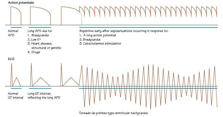
Fig. 38.2 Scheme of the mechanism of torsade-de-pointes (TDP) ventricular tachycardia. The top line represents cellular events, the bottom line those in the surface ECG. Many factors prolong the duration of the action potential (APD), some are listed, of which bradycardia on top of an already prolonged APD is important. Catecholamines don’t prolong the APD, but are important in promoting TDP. Arrhythmic APD prolongation predisposes to repetitive early after depolarizations (EADs), which may be linked to Ca2+ overload. With EADs, repeated action potentials occur, starting towards the end of the pathologically prolonged APD. These are often much shorter in duration than the original AP, and fire from a higher membrane potential. They underlie many cases of TDP: whether the entire TDP sequence is driven from a cell or group of cells with repetitive EADs, or whether self-perpetuating re-entrant circuits occur in the ventricle is unclear (probably both mechanisms apply in different patients). A burst of TDP often starts with an underlying bradycardia (which prolongs the APD), then an ectopic (a short RR interval), which gives rise to a compensatory pause (a long RR interval), which further prolongs the APD, setting of the burst of TDP (with the interval from the last normal beat to the first TDP beat being short), i.e. there is a characteristic short–long–short sequence before many episodes of TDP. Suppressing the compensatory pause and its associated arrhythmogenic APD lengthening (e.g. with a pacemaker) can sometimes prevent episodes of TDP.

Fig. 38.3 Torsade-de-pointes type ventricular tachycardia, showing classical ‘twisting of the points’.

Psychological disease and particularly its treatment can have a number of ECG manifestations.
Psychological stress
Stay updated, free articles. Join our Telegram channel

Full access? Get Clinical Tree


