Chapter 15 Principles of Imaging in Vascular Disease
Magnetic Resonance Angiography
Time-of-Flight Angiography
Time-of-flight angiography is performed using a flow-compensated gradient refocused sequence. Data are typically acquired as a stack of two-dimensional slices or as a single three-dimensional volume. In this technique, stationary tissues in the slice or volume of interest are saturated from repeated radiofrequency pulses and have low signal intensity.1–3 Blood flowing into the imaging has not been subjected to these radiofrequency pulses and is therefore fully magnetized (unsaturated). When it flows into the volume, it is bright compared with the stationary background tissues. This inflow technique works well in relatively normal arteries and veins, such as carotid arteries, the cerebral vasculature, vessels of the feet, the inferior vena cava, and iliac veins. Selective saturation pulses can be applied, allowing selective saturation of blood entering the imaging volume, such that either the arterial or the venous signal can be suppressed. With this technique, separate images of the arterial and venous system can be acquired.
A limitation of this technique is in-plane saturation, which results in signal dropout when the long axis of the vessel coincides with the scan plane or in the presence of slowly flowing blood in tortuous arteries. Another limitation is turbulence-induced signal loss in and distal to a stenosis.4,5 In addition, the relatively long echo times required for gradient moment nulling make time-of-flight imaging sensitive to susceptibility artifacts from bowel gas, implanted metallic objects (e.g., clips), or other air-tissue interfaces. These artifacts are a primary reason for the inaccuracy of time-of-flight MRA. In addition, imaging times are lengthy because vessels need to be imaged perpendicular to the long axis of the vessel and a stack of slices must be acquired. Long acquisition times can lead to artifacts caused by slice misregistration or patient motion.
Phase-Contrast Angiography
Phase-contrast angiography uses velocity-induced phase shifts, which occur as blood flows through a magnetic field in the presence of flow-encoding gradients.1,6,7 Two images created with opposite bipolar flow-encoding gradients are subtracted from each other; in the phase difference image, the residual phase is proportional to velocity. Stationary tissues do not undergo a velocity-induced phase shift in either image; they are suppressed and therefore subtract completely. The flow-encoding gradients can be in any direction or in multiple directions, depending on the selected flow sensitivity. Phase-contrast angiography can be performed as a two- or three-dimensional gradient refocused sequence and is improved after contrast administration.8 It has been used in the evaluation of the renal arteries, carotid arteries, and portal veins. It is also used to measure flow velocity. When used with cardiac gating, a time-resolved velocity profile can be generated, providing quantitative measurement of the flow rate.
There are limitations to the technique. The amplitude of the preselected bipolar gradient determines the degree of velocity encoding (VENC). Phase-contrast sequences encode only a specified range of velocities.9 In occlusive disease, turbulence causes a wide spectrum of rapidly changing velocities, which produce intravoxel phase dispersion and signal loss. Artifactual signal loss at sites of vessel stenosis is common. The sequence is also susceptible to measurement degradation from cardiac, respiratory, and translational motion. Background subtraction can be problematic. Currently, because faster MR angiographic techniques are available, phase contrast techniques are used primarily for the measurement of flow velocity.
Three-Dimensional Contrast-Enhanced Magnetic Resonance Angiography
Three-dimensional time-of-flight gadolinium-enhanced MRA is the most widely used and useful of the MRA techniques. It does not rely on motion of blood to create flow signal. A contrast agent, typically gadolinium chelate, is given to shorten the T1 (spin-lattice) relaxation time of blood so that it is significantly shorter than that of surrounding tissues. Blood is imaged directly using a T1-weighted sequence. Blood is bright relative to background tissues during the arterial phase of three-dimensional contrast-enhanced MRA, because contrast transiently reduces the arterial blood T1 to less than that of the brightest background tissue (fat). This technique reduces the sensitivity to turbulence, and in-plane saturation effects are eliminated.4,5 With this technique, a number of slices are oriented in the plane of the target vessels, permitting rapid imaging of a large field of view covering a large region of the vascular system. Contrast-enhanced MRA is fast and affords high-quality breath-hold and non–breath-hold angiograms. Dynamic contrast-enhanced MRA exploits the transient shortening in blood T1 after the intravenous administration of a contrast agent using a fast, three-dimensional spoiled gradient echo sequence. This is a first-pass technique, because the contrast agents currently used are extracellular agents and the gadolinium chelate rapidly leaks into the extravascular space. However, repeated slice volumes over the region of interest can be acquired during passage of the contrast allowing images in both the arterial and venous phases. Typical repetition times for contrast-enhanced MRA are less than 5 msec, with echo times of 1 to 2 msec and total scan times of 10 to 30 seconds. The sequences that use T1-weighted gradient echo, T1 fast field echo, or fast low-angle shot have high spatial resolution and a high signal-to-noise ratio. The images can be acquired in a breath-hold fashion and reformatted in any plane.10–14 Because intravascular signal is dependent on T1 relaxation rather than inflow or phase accumulation, in-plane saturation and signal loss owing to turbulence are not significant.4
During the first-pass arterial phase, imaging is done before vascular contrast equilibration. Using a first-pass technique, steady-state background signal is nearly eliminated. Once the contrast is injected and dynamic imaging is done, however, a second bolus injection of contrast material must be given to repeat the process over, for example, a second region of interest. When the second dose is given, residual soft tissue enhancement can obscure vascular detail.15 Subtraction techniques, using a precontrast mask, are typically used to remove background tissues. When the second or third dose of contrast is used, subtraction is mandatory. Timing is important in contrast-enhanced MRA. Unlike CT, MRA does not map spatial data linearly over time.16 With three-dimensional MRI, all the three-dimensional Fourier or K-space data (the information from which the image is constructed) are collected before individual slices are reconstructed. K-space maps spatial frequencies rather than spatial data. Consequently, K-space data do not correspond to image space directly. Different portions of K-space determine the features of the image. The center of K-space, which records low spatial frequencies, affects contrast, whereas the periphery of K-space records high spatial frequencies, which contribute to the fine details, such as edges.16,17
Intravascular T1 signal intensity is determined by the gadolinium concentration at the time the center of K-space is collected.17 The timing is synchronized so that the midportion of the bolus arrives at the desired site as the center of K-space is being collected. Perfect timing produces maximal arterial signal with minimal venous signal. If central K-space data are acquired too early, while arterial gadolinium is increasing rapidly, ringing or banding artifacts may be generated. Acquiring central K-space data too late leads to reduced arterial signal intensity and enhancement of venous structures. Several methods have been used to obtain proper bolus timing, including simple estimates of the travel time of the bolus from the site of injection to the region of interest. For example, in a healthy patient, the travel time from the antecubital vein to the aorta is approximately 15 seconds; in a patient with cardiac disease or an aneurysm, it is 25 to 35 seconds. To estimate contrast travel time more precisely, a test bolus can be used. In this technique, 1 to 2 mL of contrast followed by a 10- to 15-mL saline flush is injected at the same rate as the planned injection. Multiple single-slice fast gradient echo images of the appropriate vascular regions are then obtained as rapidly as possible—typically, every 1 to 2 seconds for a given period—and the time to peak enhancement (contrast travel time) is determined in the region of interest. A limitation of this technique is the setup time; also, the redistribution of the test bolus to the interstitial space may add to the background signal. Other timing techniques used include MR fluoroscopy.18 In this technique, two two-dimensional sagittal gradient refocused images are obtained rapidly (<1 second per image) throughout the region of interest. Images are generated in near real time and updated at a rate greater than one image every second. The bolus time is watched, and when it arrives, the operator switches over to the three-dimensional MRA sequence. This technique may be helpful in cases of asymmetrical flow owing to asymmetrical stenoses.
Another technique is a temporally resolved method in which multiple three-dimensional data sets are rapidly acquired (over 2 to 8 seconds) without any predetermined timing; injection and scanning are begun simultaneously.19–23 The operator then selects the desired image set. Other techniques used to facilitate MRA involve simply scanning faster using parallel imaging techniques and using alternative K-space acquisition techniques.
Respiratory motion causes image blurring, ghosting, and signal loss. In three-dimensional imaging, blurring occurs in the direction of motion, whereas ghosting is more pronounced in the phase-encoding direction.24 Before the availability of fast imaging systems, MRA techniques were too long to permit breath holding. Breath holding results in improved images in abdominal and thoracic MRA and facilitates the visualization of small vessels, such as the renal arteries.25 It does not appear to be as critical in the evaluation of the carotids. Most ambulatory patients can hold their breath 20 to 30 seconds, and imaging is usually done on inspiration.
Gadolinium-Based Contrast Agents
Gadolinium decreases both spin-lattice (T1) and spin-spin (T2) relaxation times. Because gadolinium is toxic in its natural form, it is chelated with ligands, such as gadopentetate dimeglumine, gadoteridol, or gadodiamide, to form MR contrast agents.26 These agents are extracellular and pass from the intravascular compartment into the interstitial space in a matter of minutes.4,5 Measurements of T1 shortening at different cardiac outputs have shown that injection rates greater than approximately 2 mL/s do not increase T1 shortening. Signal intensity increases asymptotically as the injection rate increases, with negligible increases seen beyond a rate of 4 to 5 mL/s.27,28
From their introduction, these agents were found to have a high safety margin and a low rate of adverse effects. It has been estimated that approximately 6 million doses of gadolinium containing contrast agents are administered annually.29,30 However, in the late 1990s a new disease was described, and in 2006 was shown to be associated with the use of gadolinium containing contrast agents.31 This disease, nephrogenic systemic fibrosis, has a low incidence; there are only several hundred cases reported in the literature.32,33 It is characterized by fibrosis of the skin, connective tissue, and internal organs, eye changes, and flexion and extension contractures. There is currently no consistently successful treatment, and it is potentially fatal. The majority of cases have occurred in patients with end-stage renal disease, chronic kidney disease, acute kidney injury, and acute renal insufficiency of any severity owing to hepatorenal syndrome, and in patients requiring dialysis or those in the perioperative liver transplantation period. These patients and patients with glomerular filtration rate (GFR) of 30 mL/min per 1.73 m2 or less are at high risk. It is recommended that gadolinium-based contrast agents be avoided in these high-risk patients and alternative imaging methods be used unless the information cannot be obtained with non-contrast MRI or other methods. It is also recommended that all patients undergo screening for renal insufficiency before receiving gadolinium-based contrast agents and package insert doses not be exceeded.34 The risk in patients with normal renal function is unknown, and to date there have been no reported cases in patients with normal renal function.
Postprocessing Techniques
Three-dimensional contrast-enhanced MRA produces a contiguous volume of data. In most body MRA studies, the volume is asymmetrical (e.g., 400 × 300 × 64). The slice is typically viewed interactively using a computer workstation. Thin multiplanar reformatting (MPR) can be performed,1,28 and 1- to 2-mm slices can be viewed in multiple planes (axial, sagittal, and oblique).35,36
Because the thin sections do not display the entire vessel, the maximum-intensity projection (MIP) processing technique is often used.37,38 With this algorithm, the user first selects the volume or portion of the volume to be evaluated. The algorithm then generates rays perpendicular to the viewing plane, records the maximum intensity of any voxel encountered along that ray, and assigns that maximum value to the corresponding pixel in the output image. This process results in images similar in appearance to conventional angiograms.
The MIP algorithm has some limitations. A major problem occurs when stationary tissue or structures within stationary tissue have a higher signal intensity than the vessels of interest. This can occur in the presence of crossing vessels, hemorrhage, fat, metallic susceptibility artifacts, or motion artifacts. This results in mapping of these extra signals into the projection image, producing a discontinuity in vessel signal that mimics vessel stenosis.1 Reducing the thickness of the MIP subvolume to exclude as much extraneous tissue as possible can mitigate this limitation. Underestimation of vessel diameter is another limitation of the technique.
Subtraction techniques are also used to improve vessel visibility. These techniques are performed by a complex subtraction of precontrast and postcontrast raw data sets39,40 and are routinely used in contrast-enhanced MRA of the extremities.
Clinical Applications
Extracranial Carotid Arteries
The carotid arteries can be well seen with contrast-enhanced MRA (Figure 15-1). The short circulation time of the carotid circulation (4 to 6 seconds) makes timing critical; otherwise, venous overlap of the images results. Both long and short imaging times have been used to overcome this problem.
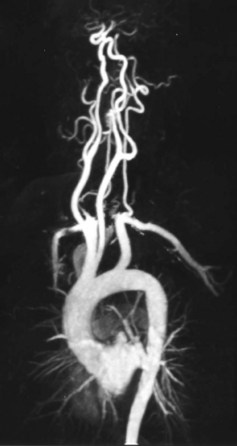
High-resolution imaging with short echo time is recommended. A major problem that impedes accurate stenosis assessment is the occurrence of high-speed turbulent jets at the site of stenosis. Specialized coils are recommended. The origins of the vessels from the aorta should be included in the imaging plane. Numerous studies have shown a high degree of correlation between stenosis measurement with contrast-enhanced MRA and digital subtraction angiography.41–46 MRI has also been used to evaluate atherosclerotic plaque in the carotid arteries.47,48
Thoracic Aorta
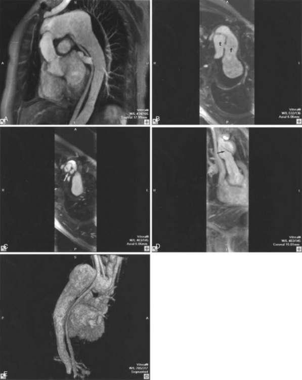
Contrast-enhanced MRA is used routinely to evaluate the thoracic aorta. It is the standard of practice for following the status of dissections and monitoring aneurysm enlargement (Figure 15-2). Imaging is usually performed in the oblique sagittal plane or coronal plane. In aortic dissection, the origins of the brachiocephalic vessels should be included. A phased array body coil is recommended when the aneurysm is confined to the thoracic aorta. The ascending, descending, and abdominal aortas are evaluated. A full evaluation of the aorta should include multiplanar cardiac-gated black blood imaging of the aortic wall to detect intramural hematoma.
Abdominal Aorta and Pelvic Vessels
Contrast-enhanced MRA is most often performed in this region to evaluate for aortic dissection or abdominal aortic aneurysm or as part of an evaluation for peripheral artery disease. The images are acquired in the coronal plane using a phased array body coil. A localizer image is obtained first in the sagittal plane, preferably with breath-holding, to determine that the entire volume of the aneurysm will be in the field of view. It is important to note that vessel calcifications are typically not seen with MRA (see Figure 15-2A).
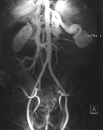
Renal Arteries
Renal contrast-enhanced MRA is performed similarly to an abdominal aortic study, although a thinner coronal slab can be used, allowing decreased slice thickness or acquisition time. High-resolution imaging is preferred. A true slice thickness of less than 2.4 mm is recommended, and breath holding is critical. Otherwise, distal renal artery branches and accessory renal arteries may be difficult to visualize. Typically, renal arteries can be seen out to the interlobar branches. Review of the raw data is critical for full assessment (Figure 15-3).
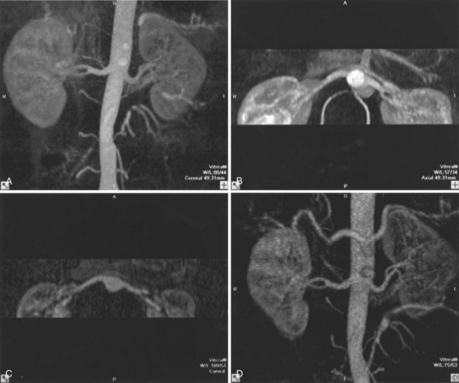
Small intrarenal branches are not seen well with current techniques. If resolution is not adequate or if motion occurs, fibromuscular dysplasia might not be detected. Examination of the source images can provide information regarding renal size and cortical thickness. A transit time can be calculated for each kidney. Renal transplant arteries can also be seen with contrast-enhanced MRA. Newer techniques that shorten acquisition time and reduce motion artifacts should result in improved visualization of the renal arteries. Although there are multiple studies describing the value of renal artery MRA, it is used largely as a screening tool in patients with acceptable GFR. In hypertensive patients, conventional arteriography is usually deferred if the renal arteries are normal. If an abnormality is detected, conventional arteriography is usually performed, at which time angioplasty or stent placement can be done (Figure 15-4). Using conventional angiography as a reference standard, the reported sensitivity and specificity of contrast-enhanced MRA for diagnosing renal artery stenosis are 88% to 100% and 70% to 100%, respectively.49
Mesenteric Vessels
The proximal portions of the mesenteric arteries are usually well seen on renal or abdominal MRA. Contrast-enhanced MRA is used often to evaluate for mesenteric ischemia caused by proximal vessel stenosis or occlusion. Evaluation of the source images and MPR are useful.50–53 Distal small vessels are usually not visualized well enough to exclude distal small vessel disease. Thin slices are recommended.
Portal Venous System
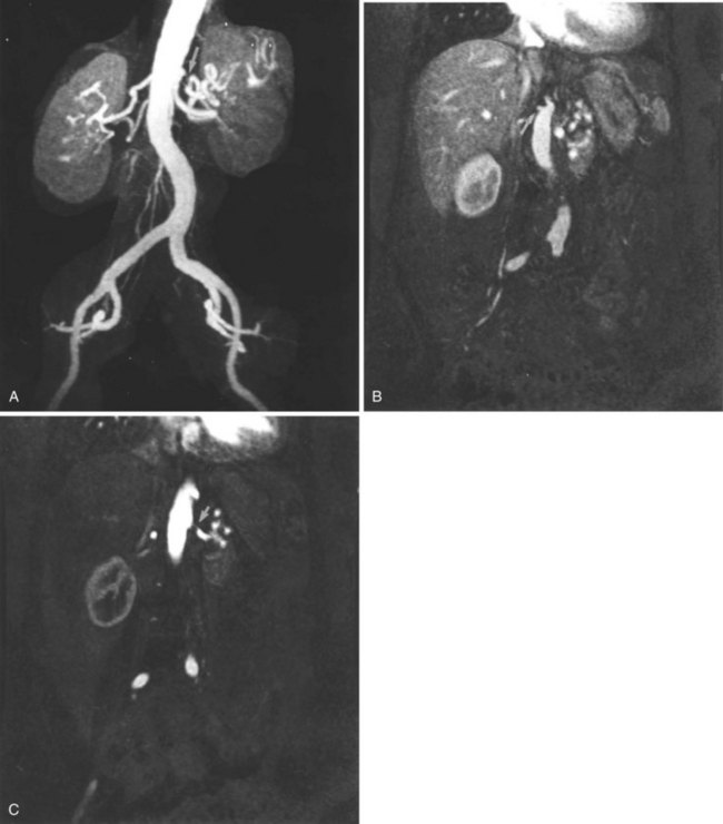
Contrast-enhanced MRA provides high-quality images of the portal system and hepatic veins. Following arterial phase images, delayed acquisitions are obtained that routinely show the portal and hepatic veins.53,54 Portal vein thrombosis and collaterals are also seen (Figure 15-5). Again, a review of the source data thin MIP subvolume and often three-dimensional volume rendering are necessary.
Peripheral Vessels
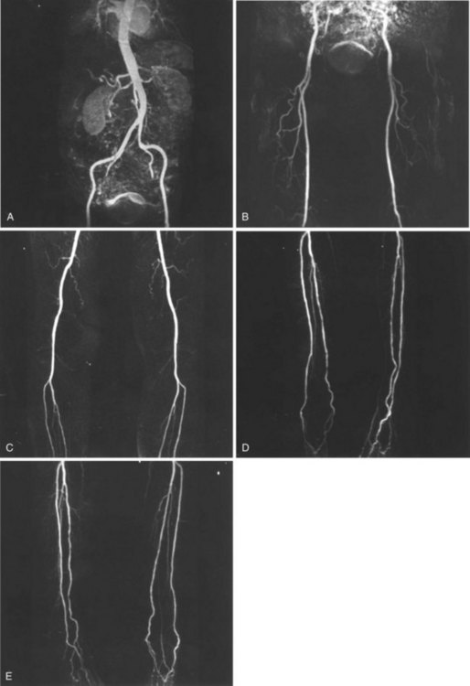
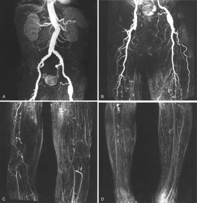
Initially, contrast-enhanced MRA of the runoff vessels of the lower extremities was limited by the restricted field of view of MR scanners.55 The field of view of most scanners, typically 400 to 500 mm, required repeated injections to cover the entire lower extremity. This imaging was typically performed by first imaging the smaller vessels of the calf, followed by repeated injections while imaging over the knee and thigh. A complete abdominal aorta and peripheral runoff study could not be performed in one setting because of soft tissue enhancement from extracellular gadolinium contrast agent. Newer scanners are equipped with a moving table, which allows image acquisition similar to that of conventional arteriography, whereby a single bolus of contrast can be followed and imaged multiple times as it travels from the aorta to the feet (Figures 15-6 to 15-8). Newer scanners also permit whole-body MRA (Figure 15-9).
< div class='tao-gold-member'>
Stay updated, free articles. Join our Telegram channel

Full access? Get Clinical Tree


