Postoperative Atelectasis


Postoperative Atelectasis


Anatomic Alterations of the Lungs
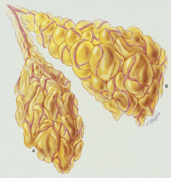

Atelectasis, in the strict sense of the term, is defined as the condition in which the lungs of the newborn remain unexpanded (airless) at birth. In the clinical setting, however, the meaning of atelectasis in all age groups generally is broadened to include partial or total collapse of previously expanded lung regions. Atelectasis may be limited to the smallest lung unit (i.e., alveolus or primary lobule*; see Figure 42-1), or it may involve an entire lung or a segment or lobe of the lung. In this chapter, postoperative atelectasis is used as a prototype of the atelectasis process.


Postoperative atelectasis commonly is seen after upper abdominal and thoracic surgical procedures.


The major pathologic and anatomic alterations associated with postoperative atelectasis include partial or total collapse of the following:


Etiology and Epidemiology


Postoperative atelectasis develops when lung expansion is decreased or when excess airway secretions cause mucous plugs, which in turn produce distal “degassing” of lung units.


Decreased Lung Expansion


Good lung expansion depends on the patient’s intact chest cage and his or her ability to generate an appropriate negative intrapleural pressure. Thoracic and upper abdominal procedures often result in a reduction in the patient’s ability to generate good lung expansion and therefore are considered as high-risk factors for subsequent development of postoperative atelectasis.


Other precipitating factors that decrease the patient’s ability to generate a negative intrapleural pressure include (1) anesthesia, (2) postoperative pain, (3) supine position, (4) obesity, (5) advanced age, (6) inadequate tidal volumes during mechanical ventilation, (7) malnutrition, (8) free fluid in the abdominal cavity (ascites), (9) diaphragmatic apraxia (e.g., topical cooling of the left phrenic nerve often occurs during cardiac surgery and may lead to an inadequate diaphragmatic movement and left lower lobe atelectasis), and (10) the presence of restrictive lung disorders (e.g., pleural effusion, pneumothorax, acute respiratory distress syndrome [ARDS], pulmonary edema, chronic interstitial lung disease, and pleural masses).


Alveolar Degassing Distal to Airway Secretions and Mucous Plugs (Airway Obstruction)


Postoperative atelectasis often is associated with retained airway secretions and mucous plugs. Precipitating factors for retained secretions include (1) decreased mucociliary transport, (2) excessive secretions, (3) inadequate hydration, (4) weak or absent cough, (5) general anesthesia, (6) smoking history, (7) gastric aspiration, and (8) certain preexisting conditions (e.g., bronchiectasis, chronic bronchitis, cystic fibrosis, asthma). When total airway obstruction develops, alveolar oxygen is absorbed into the pulmonary circulation and alveolar degassing ensues. The breathing of high oxygen concentrations favors this pathologic process.


image OVERVIEW of the Cardiopulmonary Clinical Manifestations Associated with Postoperative Atelectasis


The following clinical manifestations result from the pathologic mechanisms caused (or activated) by Atelectasis (see Figure 9-8)—the major anatomic alterations of the lungs associated with postoperative atelectasis (see Figure 42-1).


CLINICAL DATA OBTAINED AT THE PATIENT’S BEDSIDE


Jun 11, 2016 | Posted by in RESPIRATORY | Comments Off on Postoperative Atelectasis

Full access? Get Clinical Tree

Get Clinical Tree app for offline access