Fig. 6.1
Drawing of normal atrioventricular conduction tissue and concealed slowly only conducting retrograde accessory pathway supporting PJRT
Clinical and Electrocardiographic Findings
Persistent junctional reciprocating tachycardia usually presents during childhood <18 years and in approximately 50 % of patients during the first year of life. With advancing technology, the diagnosis can be entertained in utero and further evaluated using M-mode echocardiography to study the relationship of atrial and ventricular contractions in the fetus, supporting the diagnosis of PJRT. There is one familial case of PJRT where grandmother and grandson were noted to have PJRT. A 72-year-old female and in her 16-year-old grandson underwent evaluation and HLA typing. The Bw41 antigen was found in the patients as well as in the boy’s paternal uncle. This is the first documented familial case of PJRT. The possible significance and correlation with the Bw41 antigen is still under scrutiny.
Persistent junctional reciprocating tachycardia heart rates usually range from 100 to 250 beats per minute. The criteria for diagnosis of PJRT include a narrow complex tachycardia, a long R-P interval, and inverted P-waves in leads II, III, AVF, and the left lateral precordial leads (Fig. 6.2). Unusual forms of suspected PJRT have been described where similar pathway behavior supports suspected a “PJRT-like” diagnosis. In one case (see Medeiros in Suggested Reading), there was a near-incessant tachycardia, with a 1:1 atrioventricular relationship and a retrograde P wave (P)′ occurring closer to the succeeding QRS complex, i.e., with a P-R interval shorter than the RP′ interval. The tachycardia episode was characterized by alternating short and long cardiac cycles due to alternation of retrograde conduction time (RP′ interval) in a retrograde Wenckebach periodicity pattern. The authors proposed that the accessory atrioventricular connection had decremental functional properties that arborized into two atrial branches or categories with different conduction times. The fast branch initially exhibiting a 3:2 retrograde conduction block followed by a cycle length-dependent 2:1 retrograde conduction block, thereby permitting alternate use of the slow branch, which was thought to be the weakest component of the reciprocating process.
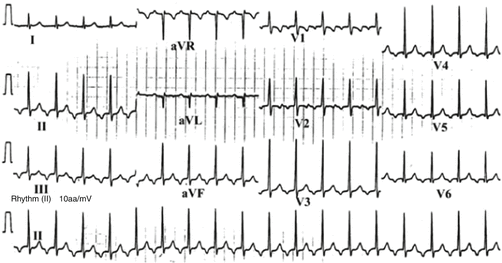

Fig. 6.2
12-Lead electrocardiogram and rhythm (bottom tracing) in a 5-year-old girl with PJRT. Note the negative P-waves in leads II, III, aVF, and the lateral precordial leads along with the long RP interval. [Reprinted from Dorostkar, P, et al., Clinical course of PJRT. Journal of the American College of Cardiology 1999;33(2):366–375. With permission from Elsevier.]
In pediatric patients with PJRT, the heart rate decreases with a PJRT cycle length increase from 200 to 300 beats per minute in the first 2 years of life, settling around heart rates of about 120–150 beats per minute as the patient matures, as can be seen in Fig. 6.3. Furthermore, as the PJRT cycle length increases the principal component appears to be associated with slowing of conduction in the concealed retrograde limb of the reentrant circuit, accounting for 64 % of the increase in the tachycardia cycle length as shown in Fig. 6.4. In contrast, the PR interval is relatively stable, accounting for only 36 % of the total increase in the tachycardia cycle length Fig. 6.5.
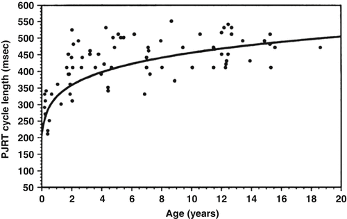
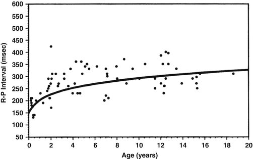
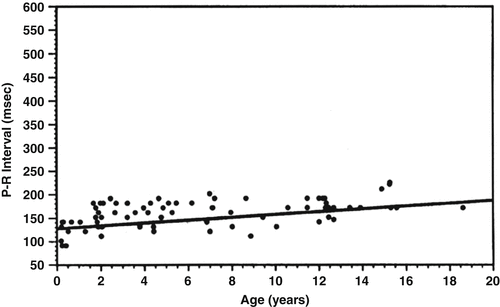

Fig. 6.3
Plot of serial tachycardia cycle length versus age years in 9 children with PJRT. Note the trend of an increase in cycle length slowing of the tachycardia with age. [Reprinted from Dorostkar, P, et al., Clinical course of PJRT. Journal of the American College of Cardiology 1999;33(2):366–375. With permission from Elsevier.]

Fig. 6.4
Plot of age versus R-P interval demonstrating a slowing in the retrograde conduction limb increased R-P interval of the reentrant circuit over time. [Reprinted from Dorostkar, P, et al., Clinical course of PJRT. Journal of the American College of Cardiology 1999;33(2):366–375. With permission from Elsevier.]

Fig. 6.5
Plot of age versus P-R interval demonstrating slow gradual increase in the P-R interval with time. [Reprinted from Dorostkar, P, et al., Clinical course of PJRT. Journal of the American College of Cardiology 1999;33(2):366–375. With permission from Elsevier.]
Although very young patients <2 years with PJRT tend to have incessant tachycardia, older subjects with slower retrograde conduction through the accessory pathway also tend to exhibit an incessant form. This apparent contradiction is most likely related to the shorter refractoriness and faster conduction velocity of all cardiac excitable tissue found in young individuals, facilitating, in the presence of the necessary anatomic substrate, i.e., another conducting pathway such as in patients with PJRT, the establishment of a fast conducting reentrant circuit. As the individual gets older and these properties lengthen, the increasingly slow retrograde conduction of the abnormal accessory pathway compensates for the longer refractoriness of the return tissues, i.e., atrium, allowing for recovery of excitability and incessant tachycardia, but at a slower rate.
Although PJRT in most patients is incessant, some subjects exhibit a paroxysmal form (Fig. 6.6). One factor that may contribute to this observation is the variation in the cardiac electrophysiologic properties between individuals and even within a single individual at different ages and physiologic states. When a longer refractory period in the atria is coupled with a faster retrograde conduction velocity in the accessory pathway, even for one cycle, the reentry circuit may be interrupted and the tachycardia transiently terminated. Clearly, the state of the autonomic nervous system and circulating catecholamines will influence the electrophysiologic properties of the circuit and vary the PJRT rate and pattern. In addition, because of the above noted variation in the electrophysiologic properties of the heart, along with the slowing of heart rate with increasing age, the arrhythmia may not be detected until later in childhood or even adulthood. Permanent junctional reciprocating tachycardia is often an asymptomatic tachycardia in an adult; however, unusual presentations have been described including presenting with complaints of syncope.


Fig. 6.6
Intermittent PJRT. S = sinus impulse; P′ = retrograde P-wave due to reciprocating PJRT impulse
Despite the increase in tachycardia cycle length with age, PJRT may continue to be incessant, and, thus, may lead to tachycardia-mediated cardiomyopathy. The age-related decrease in both the heart rate and the persistence of tachycardia may mask the diagnosis of cardiomyopathy until later in life. Some patients may present with tachycardia-related symptoms or palpitations, as well as decreased exercise tolerance, fatigue, or syncope due to an associated decreased ventricular function. Patients with PJRT may also present with clinical signs of congestive heart failure or echocardiographic findings of impaired ventricular function compatible with cardiomyopathy. Limited series of patients with PJRT have documented diminished left ventricular function in as many as 75 % of patients. Signs and symptoms of congestive heart failure may be especially evident in infants with faster PJRT heart rates, but may be absent in the adult and go undiagnosed or misdiagnosed. The tachycardia will most likely slow as the patient gets older, and some patients have infrequent, spontaneous, and intermittent periods of remission. In some patients, the ventricular shortening fraction or ejection fraction measured by echocardiogram may improve, either spontaneously with time as the tachycardia cycle length lengthens with age, or the tachycardia becomes intermittent or goes away due to successful ablation of the accessory pathway. However, multiple case reports document the benefit of ablation for control or abolition of tachycardia in patients who have incessant tachycardia-mediated myopathy. One report with a larger and older cohort of patients with PJRT, comprising 49 patients with a mean age of 43 years, noted that 16 % presented with tachycardia-induced cardiomyopathy, which was associated with a ventricular rate of about 146 ± 30 beats/min. In all cases, the myopathy regressed after successful ablation.
Electrophysiology
The tachycardia circuit traverses the normal AV nodal conduction system antegradely and returns to the atrium through a slowly conducting concealed pathway with decremental conduction properties (Fig. 6.1). The incessant nature of the tachycardia is probably due to its characteristically slow, retrograde, and unidirectional (no antegrade) conducting pathway, allowing the refractory period of the upstream cardiac tissue atria to recover so that they always present “an excitable gap” to the reciprocating impulse. The conduction velocity is typically faster and the antegrade effective refractory period of the AV node usually shorter than the conduction velocity and the retrograde effective refractory period of the accessory pathway, indicating less robust retrograde conduction. The introduction of a ventricular extrastimulus either from the right ventricular apex or the right ventricular summit during tachycardia while the His bundle is refractory, may advance, delay, or not affect the next recorded atrial electrogram and may or may not reset the tachycardia. A foreshortening of the atrial electrogram interval or termination of the tachycardia, even for only one beat, following the premature ventricular extrastimulus, strongly suggests the presence of an accessory pathway, and a delay in the next recorded atrial electrogram confirms that the slowly conducting retrograde accessory pathway is crucial to support the reentrant circuit during tachycardia. Rarely, PJRT may coexist with pre-excitation, as well as other concealed accessory pathways. In patients ≥10 years of age with shorter tachycardia cycle lengths, retrograde conduction through the accessory pathway is significantly faster in the paroxysmal form of PJRT as compared to the persistent form of PJRT.
Treatment
PJRT is often resistant to medical therapy, even with the use of one or multiple antiarrhythmic medications. A few case reports indicate limited efficacy of amiodarone therapy in patients with PJRT; one study reported that control of the incessant nature of the tachycardia was primarily related to the effects of amiodarone on the AV node rather than the accessory pathway associated with PJRT. In another cohort of patients aged 59 ± 62 months, the tachycardia was variably controlled with combinations of antiarrhythmic agents. Because of relative risks in smaller patients, deferment of definitive treatment with transcatheter ablation may be delayed until the patient is older. On the other hand, radiofrequency ablation of the accessory pathway is clearly the treatment of choice when it is judged safe to do.
Electrophysiologic Mapping and Ablation
Electrophysiologic evaluation includes assessment of retrograde conduction. The differential diagnosis of long RP tachycardias includes PJRT, atypical atrioventricular nodal reentry tachycardia, and atrial tachycardia. Delivery of a ventricular extrastimulus while the His bundle is refractory can differentiate this tachycardia from the other forms of long R-P tachycardia. The extrastimulus may advance, delay or not have any effect on the next inscribed atrial electrogram. If the atrial electrogram is delayed, the diagnosis of PJRT is confirmed. If the atrial electrogram is advanced and the tachycardia is reset, the atrioventricular reentry tachycardia and atrial tachycardia are highly unlikely. During entrainment from the ventricle, the difference between the post-pacing interval and tachycardia cycle length is shorter for patients with PJRT compared to those with atrioventricular reentry tachycardia. Ventriculo-atrial dissociation during tachycardia rules out PJRT. The slowly conducting accessory pathway of PJRT usually bridges the AV groove in the right posterior septal (inferior-septal) area where it can be mapped.
The most common site of the accessory pathway location was noted to be just superior to the coronary sinus ostium (53 %) in one series of 21 patients and right posterior septal (inferior-septal) in 25 of 32 and 28 of 37 in two other series (Fig. 6.7). Unusual locations for this pathway have been reported. The PJRT accessory pathway has been noted in the right lateral area, left posterior (inferior) and left posteroseptal (inferior-septal) regions, and even left anterior (superior) and left lateral locations. There is one case report of an epicardial location. Ablation of the earliest atrial activation during tachycardia is associated with successful ablation of the accessory pathway. Even though an accessory pathway potential is usually not noted in the target area, one case report of PJRT documents an accessory pathway potential recording at the site of successful ablation. Intuitively, it appears that ablation in smaller children might be more difficult, but there are reports of successful ablation in patients as young as infants and even pre-term neonates. With advances in technology, PJRT has been ablated successfully using remote magnetic catheter ablation techniques and non-fluoroscopic techniques. The presence of more than one pathway has also been reported with PJRT. There is one case report of antegrade conduction across the accessory pathway.
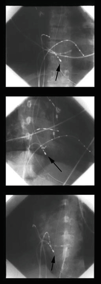

Fig. 6.7
Top: Posterior-anterior; Middle: Right anterior oblique; Bottom: Left anterior oblique radiographs during an electrophysiology study and radiofrequency ablation in a patient with PJRT. Note: the large electrode-tipped catheter arrow with the tip placed in the right infero septal area
Because the pathway is usually located in the septal area, it is thought that the risk of AV block is not insignificant, especially if a midseptal pathway is present that is anatomically closer to the normal atrioventricular node and His bundle. In such cases, one might consider the option of cryoablation. Although the success of radiofrequency ablation (Fig. 6.8) of the PJRT accessory pathway has been well documented, recurrent pathway conduction is well known. In such patients, repeat electrophysiologic study with ablation is usually successful.
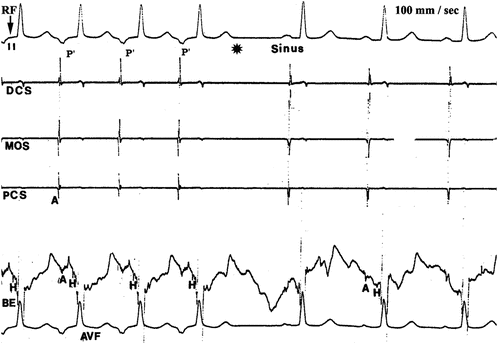

Fig. 6.8
Shortly after radiofrequency energy is started RF arrow, the PJRT is terminated by interruption of the retrograde pathway, indicated by absence asterisk of the retrograde P-wave P′
Reviewing a number of reported series, radiofrequency catheter ablation appears highly effective and safe and should be considered as the treatment of first choice in children and adult patients with PJRT. In infants, the risks and benefits of transcatheter ablation should be carefully weighed and the decision should be tailored to the specific needs of the patient.
< div class='tao-gold-member'>
Only gold members can continue reading. Log In or Register to continue
Stay updated, free articles. Join our Telegram channel

Full access? Get Clinical Tree


