Pathology of Carotid Artery Atherosclerosis


Pathology of Carotid Artery Atherosclerosis


Christopher K. Zarins and Chengpei Xu


Atherosclerosis is associated with a number of well-recognized systemic risk factors such as hyperlipidemia, hypertension, cigarette smoking, and diabetes mellitus. Atherosclerotic plaque formation is a localized rather than diffuse process, however, and preferentially affects certain segments of the arterial tree such as the carotid, coronary, and lower extremity arteries, while sparing others such as the upper extremity vessels.


Arteriosclerotic plaques in the carotid circulation usually form at the origin of the common carotid artery as it arises from the aortic arch, at the bifurcation of the common carotid artery in the neck, or intracranially in the siphon portion of the internal carotid artery. Most clinically significant carotid plaques are localized in the carotid bifurcation, making surgical treatment by endarterectomy possible. Little plaque formation occurs in the common carotid artery proximal to the bifurcation or in the internal carotid artery distal to the carotid sinus. The unique and focal pattern of plaque formation in the arterial tree at branch points and bends has been attributed to local hemodynamic conditions and to differences in artery wall susceptibility, and these have been studied most extensively at the carotid bifurcation.


Geometry of the Carotid Bifurcation


Certain geometric features of the carotid bifurcation make it particularly prone to plaque formation. It is a branching point, and as such, it is vulnerable to plaque formation, as are other branching points in the arterial tree. In addition, the carotid bifurcation is unique in that the proximal portion of the internal carotid artery is enlarged to form the carotid bulb. The cross-sectional diameter of the carotid bulb is twice that of the distal internal carotid artery, and this enlargement results in more enhanced and prominent hemodynamic alterations than are found at other branchings. The carotid bulb and the associated hemodynamic alterations might exist to permit the carotid sinus and the carotid body to carry out their functions as baroreceptors and chemoreceptors better. These functions may be particularly important early in life, but the hemodynamic conditions can predispose to plaque formation later in life.


Hemodynamic Conditions at the Carotid Bifurcation


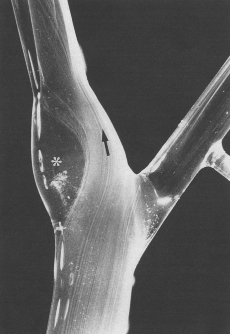
A number of flow field changes occur at arterial branch points, and these are greatly accentuated in the carotid bifurcation owing to the presence of the carotid bulb. Hydrogen bubble flow visualization studies and quantitative flow field descriptions in model carotid bifurcations have revealed that blood flow in the common carotid artery is laminar. In the carotid bifurcation, flow separates at the flow divider between the internal and external carotid branches. Flow streamlines are compressed toward the flow divider, and flow remains laminar with high velocity and wall shear stress. Along the outer wall of the widened carotid bulb there is a large area of flow separation and stasis with nonlaminar flow, low flow velocity, and low wall shear stress (Figure 1). In the region of flow separation, a reversal of axial flow and slow fluid movement upstream, with complex secondary and tertiary flow patterns with counter-rotating helical trajectories, often occurs. In the distal, tapering portion of the bulb and in the distal internal carotid artery, flow reattaches to the wall, and velocity and shear stress increase. The flow profile again becomes laminar.


Dye-washout and particle-tracking studies reveal rapid clearance along the inner wall of the internal carotid but very slow clearance from the outer sinus region, where flow separation occurs. Particles in the region of flow separation thus have an increased residence time and a greater opportunity to interact with the vessel wall. In this regard, time-dependent lipid particle vessel wall interactions would be facilitated in the region of slow flow, making plaque formation more likely to occur. In addition, bloodborne cellular elements that may be involved in atherogenesis are likely to have an increased probability of deposition on or adhesion to the vessel wall in regions of increased residence time. Radiographic and ultrasonographic studies in patients have confirmed the presence of flow separation and stasis in the outer wall region of the carotid bifurcation, where early plaques are most likely to form. In vivo human and animal studies have confirmed the relationship between low endothelial shear stress and atherosclerotic plaque localization.


The presence of the carotid bulb also results in a fluctuating change in the direction of blood flow along the outer wall of the sinus, with oscillation or change in the direction of shear stress during the cardiac pulse cycle. This shear stress oscillation has been shown to correlate strongly with early plaque formation in the carotid bifurcation. Endothelial cells normally align in the direction of flow in an overlapping arrangement. The oscillating shear stress pattern can alter the orientation of intercellular overlapping junctions and result in an increase in endothelial permeability through these junctions. Oscillation of shear stress direction also may be important because it is a systolic event and thus is directly related to the heart rate. Heart rate has been shown to be an independent risk factor for coronary atherosclerosis in humans and an important factor in coronary and carotid atherosclerosis in primates.

Stay updated, free articles. Join our Telegram channel

Aug 25, 2016 | Posted by in CARDIOLOGY | Comments Off on Pathology of Carotid Artery Atherosclerosis

Full access? Get Clinical Tree

Get Clinical Tree app for offline access