Embolic Protection Devices to Prevent Stroke during Percutaneous Angioplasty and Stenting


Embolic Protection Devices to Prevent Stroke during Percutaneous Angioplasty and Stenting


David J. O’Connor and Peter L. Faries


The use of a cerebral protection device during carotid angioplasty and stenting (CAS) has been a component of most trials demonstrating the safety and efficacy of CAS, thereby suggesting that use of cerebral protection devices is standard practice. Currently, there are two basic mechanisms to provide cerebral embolic protection: interpose a distal protection device between the lesion and the cerebral vasculature to interrupt embolic debris dislodged during angioplasty and institute proximal cessation or reversal of flow in the internal carotid artery. The disadvantage of a filter protection device is that it requires crossing the atherosclerotic lesion without protection. The major advantage of filters, however, is that they preserve antegrade flow and perfusion to the brain. This chapter focuses on the use of the distal embolic protection, including the microporous filter and temporary occlusion balloon, during carotid angioplasty and stenting.


Device Characteristics


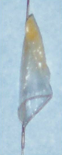
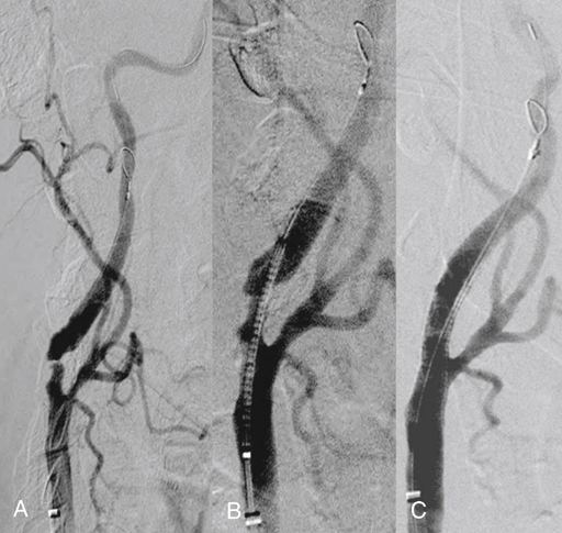
Microporous filters are designed to catch embolic debris that is liberated during CAS before it reaches the cerebral circulation. A delivery sheath constrains the filter until it is positioned in a nondiseased segment of the internal carotid artery distal to the area to be stented. Once the filter is deployed, antegrade cerebral perfusion is maintained through the filter throughout the procedure. Embolic material dislodged during balloon angioplasty becomes trapped within the filter and is removed when the filter is reconstrained at the completion of the procedure (Figure 1). Pore sizes for filters range from 100 to 165 μm, allowing capture of particulate debris larger than the pore size yet preserving blood flow through the filter.


A variety of filters are now FDA approved for clinical use in the United States and are delivered either with the filter as a component of the angiographic wire or with separate delivery over an angiographic wire already positioned into the distal internal carotid artery. Filters that are delivered attached to a fixed wire include the Angioguard XP (Cordis, Johnson & Johnson, Miami Lakes, FL), FilterWire EZ (Boston Scientific, Natick, MA), and Accunet (Abbott Laboratories, Abbott Park, IL). Emboshield NAV6 (Abbott Laboratories) and SpiderFx (EV3, Plymouth, MN) are delivered using a wire positioned independently from the filter.


The PercuSurge GuardWire (Medtronic) is a temporary occlusion balloon originally developed for use in the treatment of saphenous vein coronary artery bypass grafts. It has been adapted for use in CAS, where it prevents distal embolization by occluding blood flow in the internal carotid artery distal to the lesion. The balloon is a component of an angiographic wire and is inflated through the hypotube within the core of the wire. Similar to filter devices, the balloon is inflated in a normal segment of the internal carotid artery after passage through the lesion. An Export Catheter (Medtronic) is used to aspirate the debris and standing column of blood in the internal carotid artery between the angioplasty site and the occlusion balloon. An advantage of this device is the lower device-crossing profile and greater flexibility compared to filters; however, of necessity, cerebral perfusion is temporarily interrupted during the procedure.


Selection of Devices


There are no randomized trials comparing the advantages of one cerebral protection device against another; however, certain characteristics of patients and anatomy can help guide the choice of the best device on an individual patient basis. Particular attention should be focused on the carotid bifurcation lesion and the distal internal carotid artery. The anatomy of the access vessels, aortic arch, and common and external carotid arteries can also affect the selection of the embolic protection device, particularly if a larger-caliber proximal occlusion device is being considered. Proximal balloon occlusion should be avoided in the presence of a diseased aortic arch or proximal common carotid artery, because passage of the larger sheath and balloon inflation in the common carotid can lead to vessel damage or embolization.


Characteristics of the lesion at the carotid bifurcation are also important in selecting a device. A tight, calcified lesion can make passage of a fixed-wire filter system difficult as a result of its larger profile. Extensive tortuosity of the vessel at or immediately distal to the target lesion can also increase the difficulty of advancing a protection device that is attached to the guidewire because of the inflexibility or stiffness of the filter component. In these instances, a free-wire filter system or proximal balloon occlusion should be considered. Advancing a guidewire initially, independent from the filter, provides greater flexibility. Once the wire is positioned in the internal carotid artery distal to the target, the filter can be delivered over the wire with greater ease.


Filters and distal occlusion balloons should be deployed in a straight and nondiseased segment of the internal carotid artery that is of a sufficient distance distally to allow angioplasty and stent insertion, bearing in mind that the lead component of the balloon and stent delivery system might extend beyond the working component. Proximal balloon occlusion is an option when the internal carotid does not have a satisfactory distal landing zone.


Consideration of sufficient collateral circulation and the patient’s ability to tolerate temporary cessation of antegrade cerebral perfusion should also be a factor in selecting an embolic protection device. A microporous filter is a viable option in patients with poor collateral circulation.


Technique


Placement of a distal embolic protection device is preceded by obtaining femoral access; performing diagnostic aortic arch, carotid, and cerebral angiography; and placing a 6-Fr sheath in the common carotid artery proximal to the target lesion. Attention should be paid to the characteristics of the aortic arch, the complexity of the lesion at the carotid bifurcation, the distal internal carotid landing zone, and the presence of cerebral collateral circulation (see “Selection of Devices”). The patient is anticoagulated with intravenous heparin to an activated clotting time of 250 sec or greater.


If a fixed-wire filter system is chosen, the wire with the filter inside the delivery system is advanced past the lesion under fluoroscopic visualization into a straight segment of internal carotid artery a few centimeters distal to the proposed level of stent placement. Deployment of the filter in an arterial segment in which the walls are parallel allows optimal apposition of the filter to the luminal surface of the internal carotid artery. The delivery system is removed, resulting in deployment of the self-expanding filter. Angiography is performed to confirm proper placement, adequate wall apposition, and preservation of antegrade cerebral blood flow (Figure 2).


Stay updated, free articles. Join our Telegram channel

Aug 25, 2016 | Posted by in CARDIOLOGY | Comments Off on Embolic Protection Devices to Prevent Stroke during Percutaneous Angioplasty and Stenting

Full access? Get Clinical Tree

Get Clinical Tree app for offline access