Pathogenesis of Inflammatory Aortic Aneurysms


Pathogenesis of Inflammatory Aortic Aneurysms


Matthew T. Allemang, Kathryn S. Dyhdalo and Jerry Goldstone


Among the thousands of abdominal aortic aneurysms (AAAs) treated each year, the origin of a few are enigmatic. The inflammatory aortic aneurysm was originally described by Walker in 1972. One of the largest retrospective reviews consisted of 127 patients encountered over a 30-year period in Rochester, MN. Approximately 5% to 10% of all AAAs are of the inflammatory type. They share the same epidemiologic risk factors as the typical AAA, including male gender, smoking, and a genetic predisposition. However, inflammatory AAA occurs at a younger age and in a higher percentage of smokers, and they are more often symptomatic. The inflammatory form also shows frequent signs and symptoms of systemic inflammation with malaise, weight loss, and fever as well as an elevated erythrocyte sedimentation rate (ESR) and C-reactive protein (CRP). Although most inflammatory aortic aneurysms involve the infrarenal aorta, they can occur in other sites as well. In the series presented by Pennell and colleagues, 13.4% either were thoracic or had a thoracoabdominal component.


Pathology


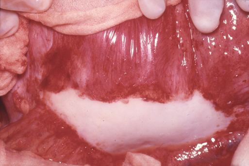
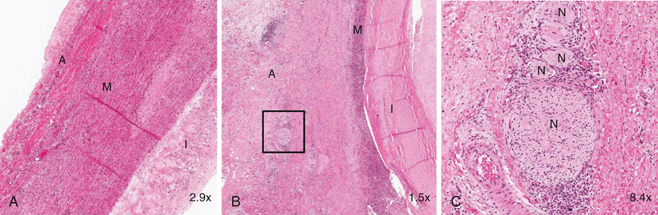
Inflammatory aneurysms are characterized by a pearly white thickened, highly vascular outer layer with dense localized adhesions to surrounding structures, most often the duodenum, inferior vena cava, left renal vein, and ureters. Any adjacent structure can be involved (Figure 1). It is these abundant unnatural bonds that make the operative approach for an inflammatory AAA more difficult than for a typical open AAA repair and has led many to suggest alternative surgical techniques such as minimal dissection of the duodenum off the aneurysm, retroperitoneal approach, or, more recently, endovascular exclusion. The aneurysmal wall thickening is predominantly located in the anterolateral locations with posterior sparing.


Microscopically, inflammatory aneurysms are similar to AAA in regard to advanced atherosclerotic changes in the intima and marked medial thinning with degeneration of elastin and loss of smooth muscle cells. The adventitial surface is where the two types of aneurysms differ. The adventitia is markedly thicker (1–4 cm) in inflammatory aneurysms, with extensive fibrosis and a chronic inflammatory infiltrate including plasma cells, histiocytes, lymphoid follicles with germinal centers, and granulomas. This inflammatory reaction also involves the medial layer, and it can contain eosinophils as well as areas of periaortic phlebitis, endarteritis of the vasa vasorum, and perineural inflammation; these changes have rarely been described in typical AAA (Figure 2). Some have noted that the histology mimics that of Takayasu’s arteritis or giant cell arteritis when taken out of clinical context. These histologic findings involving the aortic wall are continuous with identical inflammatory findings in the retroperitoneal space.


Etiology


Although inflammatory aortic aneurysms were first described more than 40 years ago, there is still much unknown about their origins. It is unclear whether inflammatory aortic aneurysms represent a distinct clinicopathologic entity or an extreme variant of the lesser inflammation that is related to the more common aneurysmal process.


A Severe Variant of Aortic Aneurysm


Although inflammatory aortic aneurysms were originally described as a discrete entity, Rose and Dent first suggested the possibility that they were severe variants of the more common atherosclerotic aneurysm. They analyzed 51 consecutive aortic aneurysmal tissue samples and graded them according to the amount of chronic inflammation. Their results showed a spectrum of inflammatory severity with no clear delineation between the aneurysms when evaluated histologically.


This concept of inflammatory aneurysms existing as a severe variant of AAA received support from the theory that many aortic aneurysms are caused by an unknown antigen that initiates an inflammatory process. These proponents point to the similarities of specific class II human leukocyte antigens (HLAs) in patients with inflammatory aortic aneurysms and those with the more typical aneurysm. Both aneurysm groups express different HLA class II alleles compared to aortic tissue from normal controls.


This unknown antigen has been hypothesized to be either a lipid or a product of lipid oxidation found in atherosclerotic plaques. The theory is that plaque macrophages present the antigen to B and T cells, causing a local inflammatory response that propagates to cause periaortic fibrosis and persistent chronic inflammation. Other investigators have suggested possible infection with cytomegalovirus or Chlamydia pneumoniae. Finally, with the higher prevalence of inflammatory aneurysms in smokers, it has been suggested that this environmental risk factor plays a direct role in the severe inflammatory reaction.

Stay updated, free articles. Join our Telegram channel

Aug 25, 2016 | Posted by in CARDIOLOGY | Comments Off on Pathogenesis of Inflammatory Aortic Aneurysms

Full access? Get Clinical Tree

Get Clinical Tree app for offline access