Pathogenesis of Abdominal Aortic Aneurysms


Pathogenesis of Abdominal Aortic Aneurysms


Sean J. English and Gilbert R. Upchurch, Jr.


Abdominal aortic aneurysm (AAA) development is a complex, multifactorial process involving destructive remodeling of aortic wall connective tissue. Four interrelated factors involved in this process include: (1) chronic inflammation associated with neovascularization and increased proinflammatory cytokine production, (2) increased and dysregulated production of matrix-degrading proteinases, (3) destruction of structural matrix proteins, and (4) decreased medial smooth muscle cell (SMC) presence, resulting in impaired connective tissue repair. This understanding has developed from a characterization of human AAA tissue, as well as the use of different animal models that replicate human disease.


Biomechanical Wall Stress and Structural Pathology


Global tension on the aortic wall is a function of many factors, including the orthogonal hydraulic conductivity of the aortic wall from the lumen to the adventitia. The associated activity through the aortic wall depends on a number of factors including blood pressure, shear stress, and filtration through the wall. This movement is likely enhanced by the porosity of the intraluminal thrombus and an associated lack of aortic endothelium at this location, decreased shear caused by recirculation vortices in blood flow, and increased pulsatility caused by elastic fiber degradation and wall dilation. As a result, pathogenic factors are directed outward through the media to the adventitia.


That AAA formation predominantly occurs in the infrarenal aorta specifically highlights structural, biologic, and mechanical differences in the aorta along its length. The number of elastin layers decreases caudally along the length of the aorta, from the ascending aorta to the iliac arteries. Approximately half as many aortic lamellae are present in the abdominal aorta compared to the thoracic aorta. This becomes clinically relevant as decreased aortic elastin predisposes the wall to dilation. This anatomic variability is reflected in a decreasing elastin-to-collagen ratio along the abdominal aorta, in a proximal to distal direction. Furthermore, given that the half-life of aortic elastin is approximately 50 years, decreased elastin content in human AAAs is likely caused by increased elastolysis, as opposed to decreased synthesis. The presence of atherosclerosis can also prevent normal elastogenesis, resulting in poorly cross-linked, immature elastin. Elastolysis can thus expose neoantigens that provoke an immune response; tropoelastin and elastin-derived peptides are known to stimulate neutrophil, fibroblast, and macrophage chemotaxis.


The tensile strength of the aorta is primarily attributed to interstitial collagen. Aortic wall collagen is made up of types I (75%) and III (25%) collagen fibers. The distribution of collagen types in AAA tissue does not appear to change, but the amount of collagen in terms of total aortic wall protein is significantly increased. This is evident when considering the increased wall stiffness of human AAAs and the tensile strength required to maintain a dilated aneurysm. There is likely a balance between degradation and repair of connective tissue, with fibroproliferative repair commonly occurring in the adventitia, limiting aortic wall dilation.


Intraluminal Thrombus


Aortic intraluminal thrombus (ILT) is composed of multiple layers and is believed to play an important role during AAA development. ILT is composed of a fresh luminal layer with cross-linked fibrin, as well as an abluminal, actively fibrinolysing layer. Repetitive luminal clotting and abluminal lysis over time generate a network of canaliculi, which allows further macromolecular trafficking. Red blood cell (RBC) hemagglutination occurs at the luminal layer, resulting in the release of free hemoglobin, with associated fibrin formation platelet and thrombin activation. RBCs are degraded in the adjacent layer, with pro-oxidant hemoglobin released, ultimately taking part in other chemical reactions and resulting in the release of free radical species that damage adjacent tissue. The involvement of nicotinamide adenine dinucleotide phosphate (NADPH) oxidase, myeloperoxidase, and catalytic iron all appear to play a role in this process. Antioxidant enzymatic activity of glutathione reductase, glutathione peroxidase, and superoxide dismutase are reduced in AAA tissue compared to that of control tissue.


Leukocyte presence is maintained to a greater extent in the luminal layer than that observed in the intervening and abluminal layers. Leukoyctes release a number of enzymes, including serine proteases (i.e., urokinase-type plasminogen activator [uPA]), elastase, cathepsins, and matrix metalloproteinases (MMPs) 9 and 8. Neutrophils are 12 times more prevalent in the ILT than in the circulating blood, and this luminal presence is associated with increased levels of MMP8 and MMP9, as well as elastase. A balance of coagulation and fibrinolysis is established by neutrophils, with both enhanced cleavage of tissue factor plasma inhibitor and fibrin. The presence of neutrophils in the ILT prevents colonization of the clot by mesenchymal progenitor cells, delaying aortic wall healing.


Tissue plasminogen activator (tPA) and plasminogen are retained at the luminal layer of the ILT, owing to the ability of these molecules to bind free lysine residues in the fibrin polymer, resulting in delayed fibrinolysis to allow continued coagulation. However, the greatest degree of fibrinolysis occurs at the abluminal surface between the ILT and the aortic wall. uPA, released by neutrophils, is also present in this luminal layer. Plasminogen is converted to plasmin by a variety of enzymes, including tPA and uPA, and in its active form, plasmin activates MMPs, mobilizes transforming growth factor-β (TGF-β), and degrades surrounding proteins.


Matrix Metalloproteinases and Endogenous Proteases


MMPs play an integral role in the inflammatory processes associated with AAA formation (Figure 1). The MMPs include at least 15 structurally related proteinases that are a subfamily of the metazincin superfamily of proteinases. Substrates of the MMPs include elastin, noncollagenase extracellular matrix (ECM) proteins, including fibronectin and laminin, and nonstructural components. The nonstructural components include interleukin (IL)-1α, IL-1β, IL-2 receptor, active MMP9, chemokine ligand 5, TGF-β, and several of the pro-MMPs. Four MMPs exhibit specific activity with elastin, including MMPs 2, 7, 9, and 12. It has been proposed that as the ECM is digested, signals in the form of cryptic fragments may be released, including arrestin, deprellin, endostatin, restin, elastin degradation products, and growth factors TGF-β and vascular endothelial growth factor (VEGF).
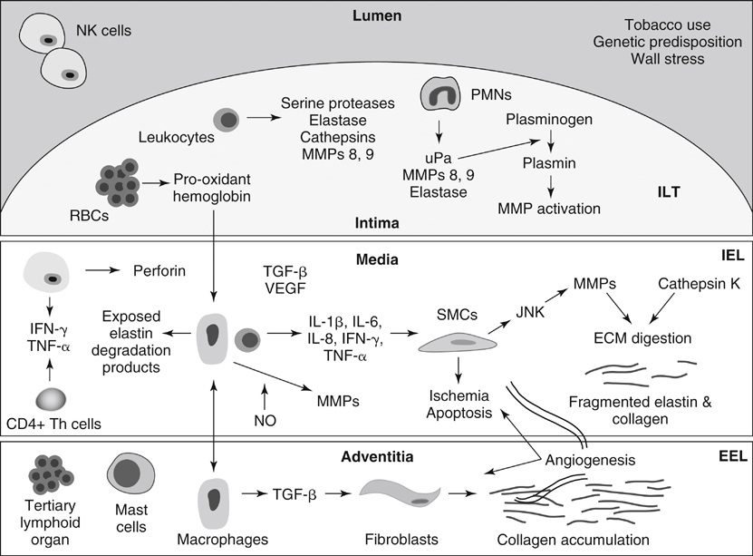


Numerous MMPS including 1, 2, 3, 8, 9, 10, 12, and 13 play roles in AAA formation. MMPs 1, 8, and 13 are capable of initiating the degradation of fibrillar collagen, and MMPs 2 and 9, both overexpressed in human and experimental AAAs, have both elastolytic and collagenolytic properties. The presence of constitutive MMP2 in smaller AAAs suggests a possible role for MMP2 in early aneurysm formation. MMP9 is not typically produced in normal aorta, but it is present in atherosclerotic plaques, suggesting a possible role in plaque rupture. MMP9 expression is increased in the serum and aortic tissue of AAA patients compared to that of patients with aortoiliac occlusive disease. There is a correlation between MMP9 expression and AAA size, and it appears to play a role in AAA expansion and ultimate rupture. Furthermore, endovascular exclusion of AAAs results in decreased MMP9 levels. The expression of MMP12 is also increased in AAA tissue, and an association with aortic media elastin has been identified.


In terms of experimental animal models, MMP9 knockout mice do not form AAAs; however, after undergoing wild-type bone marrow transplantation, the same MMP9 knockout mice exhibit an aneurysm phenotype. Secreted tissue inhibitors of metalloproteinases (TIMPs) 1, 2, and 3, as well as plasma-derived α-1-macroglobulin, cause direct inhibition of MMPs. Inflammatory states associated with increased MMP expression typically result in increased TIMP1 levels. Inhibition of the MMPs with exogenous TIMPs and α-2-macroglobulin have attenuated the development of AAAs in animal models. It has been demonstrated that doxycycline, a tetracycline antibiotic, is a nonselective MMP inhibitor, and nonantibiotic tetracycline derivatives have demonstrated MMP inhibition and have been used to prevent AAA formation in animal models and in small human studies.


The cystine proteases, including cathepsins K, L, and S, also play roles in AAA formation. The expression of all cathepsins is increased in human AAA tissue compared to normal aortic tissue. Cathepsin K is the most potent elastolytic enzyme. Cystatin C plays the greatest inhibitory role against the cathepsins, and it is ubiquitously expressed throughout the human body. The constitutive expression of cystatin C is significantly decreased in AAA patients. Furthermore, immunohistochemical analyses of human AAA tissue have demonstrated increases in cathepsin S expression and decreases in cystatin C expression compared to that in normal AAA tissue. Cathepsin S–deficient mice demonstrate less aortic dilation after elastase exposure compared to wild-type mice.

Stay updated, free articles. Join our Telegram channel

Aug 25, 2016 | Posted by in CARDIOLOGY | Comments Off on Pathogenesis of Abdominal Aortic Aneurysms

Full access? Get Clinical Tree

Get Clinical Tree app for offline access