Patent Ductus Arteriosus
Incision
The ductus arteriosus can be adequately exposed through a small left anterior thoracotomy. A limited left posterolateral thoracotomy through the fourth intercostal space partially dividing the latissimus dorsi muscle and preserving the serratus anterior muscle provides good exposure and is more commonly used. The skin incision can be quite short, especially in premature infants.
Surgical Anatomy
The ductus arteriosus runs parallel to the aortic arch from the superior aspect of the origin of the left pulmonary artery and passes through the pericardium to join the medial margin of the aorta at an acute angle just opposite the origin of the left subclavian artery (Fig. 14-1). The left vagus trunk enters the thorax from the root of the neck in a groove between the left subclavian artery and the left common carotid artery, crosses the aortic arch and the ductus arteriosus, and continues downward. The recurrent laryngeal branch curves around the ductus arteriosus and extends back upward into the neck. The vagus nerve gives rise to many other small branches that are important tributaries to the pulmonary and cardiac plexuses. There are usually some lymph nodes buried in the hilum of the left lung that sometimes extend upward near the inferior margin of the ductus arteriosus. The left phrenic nerve enters the thorax medial to the vagus nerve and continues downward on the pericardium.
Technique for Exposing and Dissecting the Ductus Arteriosus
The left lung is retracted inferiorly to expose the ductus arteriosus. The parietal pleura is divided longitudinally behind the vagus nerve if the intention is to retract the vagus nerve medially. Alternatively, a pleural incision may be made between the vagus and phrenic nerves when the vagus nerve is to be retracted laterally (Fig. 14-1). The incision of choice is extended superiorly along the left subclavian artery and inferiorly to the left hilum. The pleural edges are then suspended.
In an infant, the ductus is exposed by sharp dissection with scissors both from above and below. A blunt right-angled clamp is then carefully passed around the ductus to create a plane for its ligation or division. The ductus can also be occluded by the application of a metal clip.
To facilitate dissection and exposure of the posterior aspect of the ductus arteriosus, many surgeons prefer that the vagus nerve and its recurrent laryngeal branch be reflected medially on the pleural flap (Fig. 14-2). The surgeon should be aware that traction of the nerve toward the pulmonary artery causes the recurrent nerve to lie along a diagonal course behind the ductus arteriosus. Therefore, care must be taken to ensure that the recurrent nerve is not injured during dissection. Alternatively, the vagus nerve and its branches can be isolated and retracted laterally to ensure their protection during the process of dissection of the posterior wall of the ductus.
Special care should be taken when dissecting near the angle between the pulmonary artery and the ductus arteriosus because the ductus is particularly susceptible to injury. A lappet of pericardium usually covers the ductus anteriorly. It should be dissected free to ensure complete exposure of the ductus (Fig. 14-3).
The aorta is also dissected free, avoiding injury to the intercostal arteries. The posterior aspect of the ductus arteriosus is always adherent to the surrounding tissues and can be torn during the process of mobilization. Exposing the posterior aspect of the aorta and the ductus arteriosus can be facilitated by retracting the aorta medially with an atraumatic forceps or umbilical tape or vessel loop passed around the aorta (Fig. 14-4). This allows cautious dissection under direct vision.
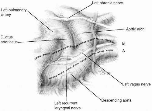 FIG 14-1. Surgical anatomy of the ductus arteriosus. A: This incision line is used if the vagus nerve is to be retracted medially. B: This incision line is used if the vagus nerve is to be retracted laterally. |
Technique for Dividing and Ligating the Ductus Arteriosus
The vagus and recurrent laryngeal nerves are identified so that they are not divided inadvertently. Two heavy Ethibond sutures are individually passed behind the ductus, which is then securely ligated (Fig. 14-5). A finer suture material may cut through the friable ductus arteriosus wall and result in hemorrhage. A purse-string suture of 4-0 Prolene may be placed between the ligatures to secure complete occlusion of the ductus (Fig. 14-5, inset).
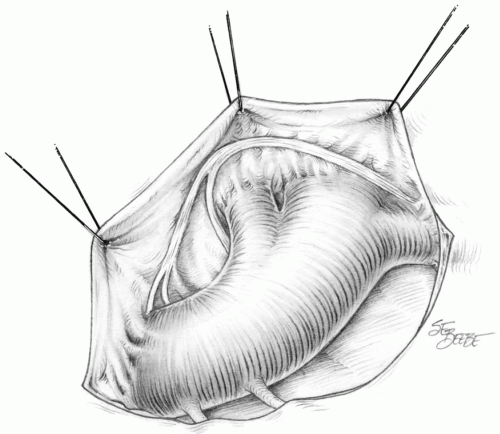 FIG 14-2. Medial reflection of the vagus nerve on the pleural flap. |
Alternatively, the ductus is divided between clamps and oversewn with fine, nonabsorbable sutures (Fig. 14-6). This technique is particularly useful when the ductus is exceptionally short and large. Another option is to occlude the ductus with one or two metal clips. This latter technique is especially applicable to premature infants.
Occasionally the posterior wall of the ductus arteriosus may be difficult to dissect, particularly in older patients. Vessel loops or umbilical tapes passed around the aorta above and below the ductus can be used to retract the aorta and dissect posteriorly until the upper loop or tape can be passed behind the aorta to encircle the ductus (Fig. 14-7).
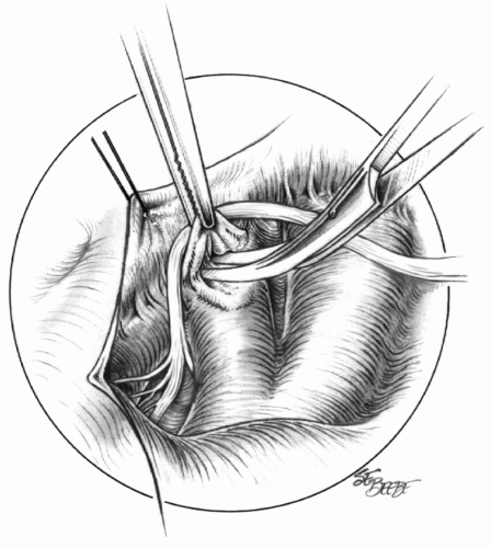 FIG 14-3. Dissecting the lappet of pericardium to ensure complete exposure of the ductus. |
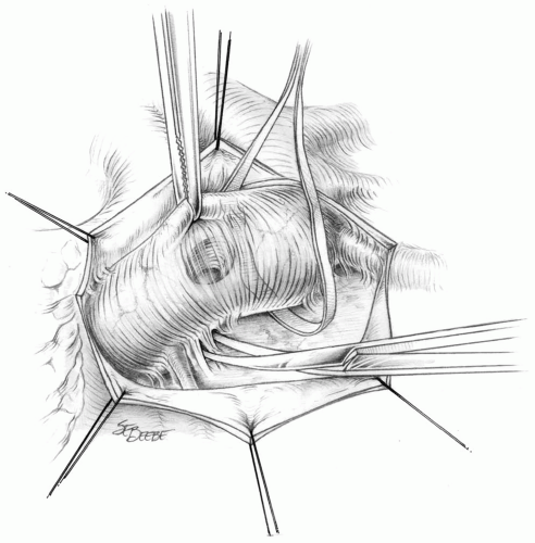 FIG 14-4. Dissecting free the aorta. |
Stay updated, free articles. Join our Telegram channel

Full access? Get Clinical Tree


