Open Surgical Treatment of Fibromuscular Dysplasia of the Carotid Artery


Open Surgical Treatment of Fibromuscular Dysplasia of the Carotid Artery


James C. Stanley


Most patients with carotid artery fibrodysplasia are likely to be asymptomatic, although the number reported without symptoms is small because most reported series are surgical experiences encompassing more advanced disease. Complications of this disease include embolization, dissections, and rupture with formation of an arteriovenous fistula. The most catastrophic clinical complication is stroke, and prophylactic interventions in asymptomatic patients are directed at reducing or eliminating this sequela of the disease. The risk of stroke or other related neurologic events varies widely and is unpredictable. Once clinical manifestations of this entity have arisen, operative intervention appears justified.


Operative Techniques


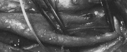
Open surgical therapy for extracranial internal carotid artery fibrodysplasia includes resection of the diseased vessel with interposition grafting, angioplasty with patch grafts for focal lesions, and graduated intraluminal dilation. Operative exposure of the carotid vessel in these cases usually requires dissection of the entire internal carotid artery to within a few centimeters of the base of the skull (Figure 1). Care must be used not to cause injury to cranial nerves IX, X, XI, and XII.


Extended exposure of the internal carotid artery at the upper cervical levels may be facilitated by subluxation of the mandible. In the author’s experience, intermittent traction on the mandible, with an external clamp inserted into the angle of the mandible, facilitates the subluxation. Others have advocated fixed traction with placement of arch bars, as well as transection of the mandibular ramus.


Patients undergoing open procedures are placed on preoperative antiplatelet agents. They are systemically anticoagulated intraoperatively with intravenous heparin before the carotid vessels are occluded, and unless incessant bleeding is present after restoring antegrade carotid flow, the heparin effect is not reversed.


Open Dilation


Graduated intraluminal dilation of the diseased internal carotid artery is usually accomplished by advancing rigid olive-tip dilators through an arteriotomy placed in the proximal carotid bulb. These are then passed the full length of the internal carotid artery to the base of the skull. Initial dilators range in diameter from 1.5 to 2.5 mm, with subsequent passage of increasingly larger dilators up to a maximum of 5.0 to 5.5 mm in diameter. Use of larger dilators should be avoided, in that they are likely to cause deep dissections within the vessel wall. Balloon catheters have been preferred by some. Because of a balloon’s radial dilation, they may be less likely to cause a dissection of the dysplastic artery when compared with longitudinal dilation by metal devices. Arterial perforation is a rare complication, but such can accompany overzealous dilation. It is important to allow back bleeding as the dilation proceeds, as a means of flushing out any debris that arises as the dysplastic septa are fractured.


Completion arteriography is not routinely performed, but if concerns arise regarding the adequacy of the dilation, then arteriography should be undertaken. Intraoperative duplex sonography may be useful, but the high cervical regions are usually difficult to insonate, and this technology is often wanting in these circumstances. Patients subjected to dilation should receive antiplatelet agents for 3 months postoperatively, until the dysplastic arterial segment has been completely remodeled.

Stay updated, free articles. Join our Telegram channel

Aug 25, 2016 | Posted by in CARDIOLOGY | Comments Off on Open Surgical Treatment of Fibromuscular Dysplasia of the Carotid Artery

Full access? Get Clinical Tree

Get Clinical Tree app for offline access