Novel Hypertrophic Cardiomyopathy Phenotype: Segmental Hypertrophy Isolated to the Posterobasal Left Ventricular Free Wall


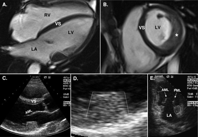
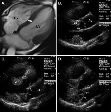
Few other diseases show the degree of phenotypic heterogeneity expressed by HC. The two novel patients reported here with isolated posterobasal LV free wall hypertrophy (and mitral valve prolapse) extend this morphologic diversity even farther, now 3 decades after the introduction of contemporary 2-dimensional imaging.


Hypertrophic cardiomyopathy (HC) is characterized by extreme diversity with respect to the distribution of left ventricular (LV) hypertrophy, identified by 2-dimensional echocardiography and cardiovascular magnetic resonance. Indeed, myriad patterns of LV wall thickening have been recognized, ranging from massive and diffuse to segmental and relatively mild. When hypertrophy is confined to a small area of the left ventricle, the site is usually in the basal anterior ventricular septum but also has been reported in the apex, anterolateral free wall, or posterior (inferior) portion of the septum.


Case Reports


We have recently identified 2 extraordinary patients with nonobstructive HC and segmental LV hypertrophy limited to the posterobasal free wall, a pattern virtually unrecognized within the broad clinical spectrum of HC ( Figures 1 and 2 ). Notably, the 2 patients also had mitral valve prolapse with redundant leaflets consistent with myxomatous degeneration ( Figure 2 ).


Figure 1


Patient 1: segmental posterobasal LV hypertrophy, the rarest wall thickness pattern in HC. Images from a 41-year-old woman with nonobstructive HC. (A,B) Cardiovascular magnetic resonance (CMR). (A) Horizontal long-axis cross-sectional image shows an area of circumscribed wall thickening in the most basal portion of the posterior LV free wall, measuring 20 mm in thickness (white dotted line) . Ventricular septum (VS) and the remainder of the LV free wall are of normal thickness and uninvolved in the hypertrophic process. (B) Basal short-axis view transecting the area of segmental hypertrophy (asterisk) . (C,D) Two-dimensional echocardiography. (C) Parasternal long-axis view shows segmental posterobasal LV hypertrophy identical in appearance to CMR image (white dotted line) , associated with nondilated left ventricle and elongated anterior mitral leaflet (AML). (D) Magnification of the area of LV wall thickening shown in (C) . (E) Posterobasal LV segmental hypertrophy seen in apical 4-chamber view (white dotted line) . LA = left atrium; LV = LV cavity; PML = posterior mitral leaflet; RV = right ventricle.


Figure 2


Patient 2: Posterobasal LV hypertrophy and associated mitral valve prolapse. Images from a 30-year-old woman with nonobstructive HC. (A) Cardiovascular magnetic resonance horizontal long-axis image shows segmental thickening of LV free wall (white broken line) . (B) Two-dimensional echocardiography at end-diastole showing the area of hypertrophy, situated posterior to the mitral valve (white broken line) . (C,D) In systole, redundant anterior and posterior mitral leaflets protrude into left atrium (LA) (arrows) , unassociated with mitral regurgitation. Ao = aorta; RV = right ventricle; VS = ventricular septum.

Only gold members can continue reading. Log In or Register to continue

Stay updated, free articles. Join our Telegram channel

Dec 22, 2016 | Posted by in CARDIOLOGY | Comments Off on Novel Hypertrophic Cardiomyopathy Phenotype: Segmental Hypertrophy Isolated to the Posterobasal Left Ventricular Free Wall

Full access? Get Clinical Tree

Get Clinical Tree app for offline access