Fig. 29.1
Raynaud’s attack in a 46-year-old female
Patients with Raynaud’s syndrome are divided into two groups. Raynaud’s disease is a term used to describe an idiopathic benign form of intermittent digital ischemia. “Raynaud’s phenomenon” describes a similar symptom complex in association with an underlying disease. An underlying condition will be discovered in association with Raynaud’s syndrome in only 5% of cases [1]. However, it is well recognized associated diseases may not be diagnosed when a patient presents with Raynaud’s attacks [2]. Distinction between Raynaud’s disease and Raynaud’s phenomenon is therefore artificial. Raynaud’s attacks should be considered a symptom that requires a diagnosis rather than a diagnosis in itself. That diagnosis in most cases will be idiopathic episodic vasospasm. In cool, damp climates, 6–20% of the population, particularly young females, report Raynaud’s symptoms [3]. Vasospasm, outside that induced by vasoactive medications or ergot, rarely results in ulceration or gangrene (Fig. 29.2). Digital ulceration or gangrene in association with Raynaud’s attacks should trigger a work-up to evaluate for digital artery occlusions and an underlying condition that leads to digital artery occlusion.
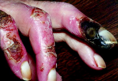
Fig. 29.2
Digital gangrene in a patient with long-standing diabetes and renal failure. Digital gangrene is almost associated with digital artery obstruction
Pathophysiology
A useful classification scheme for diagnosis of upper extremity ischemia based on vascular laboratory and physical examination findings is to divide patients into those with large artery disease and those with small artery disease. Serologic testing further facilitates determination of prognosis [4].
Small artery occlusive disease accounts for the very large majority of patients presenting with upper extremity ulceration or gangrene (Table 29.1). Large artery diseases generally produce serious upper extremity symptoms by embolization to the digital arteries (Table 29.2). Proximal occlusive disease of the subclavian artery rarely causes limb-threatening ischemia unless complicated by distal embolization to forearm or digital arteries.
Table 29.1
Diseases associated with intrinsic digital artery occlusions
Disease type | Example |
|---|---|
Connective tissue disease | Scleroderma, CREST syndrome, systemic lupus erythematosus, rheumatoid arthritis, Sjøgren’s syndrome, mixed connective tissue disease, dermatomyositis, small and medium vessel vasculitis |
Atherosclerosis and occlusive arterial disease | Atherosclerosis obliterans, atheroembolism, diabetic distal arterial disease, thromboangiitis obliterans (Buerger’s disease) |
Thromboembolism | Cardiac embolism, arterial embolism, paradoxical embolism |
Large vessel vasculitis | Takayasu’s arteritis, extracranial temporal arteritis |
Dynamic entrapment | Arterial thoracic outlet syndrome |
Occupational arterial trauma | Hypothenar hammer syndrome, vibration-induced Raynaud’s syndrome |
Drug-induced vasospasm | β-blockers, vasopressors, epinephrine, ergot, cocaine, amphetamines, vinblastine/bleomycin |
Infections | Parvovirus, hepatitis B and C, antigenemia, sepsis/disseminated intravascular coagulation |
Malignancy | Multiple myeloma, leukemia, adenocarcinoma, astrocytoma |
Hematologic | Polycythemia vera, thrombocytosis, cold agglutinins, cryoglobulinemia |
Table 29.2
Conditions resulting in embolization to upper extremity digital arteries
Source | Example |
|---|---|
Cardiac | Atrial fibrillation, atrial thrombus, valvular disease, myxoma |
Trauma | Blunt and penetrating trauma, occupational/hypothenar hammer syndrome |
Aneurysm | Subclavian artery or more peripheral artery aneurysm (ulnar artery aneurysm) |
Fibromuscular disease |
Patients with vasospastic Raynaud’s syndrome do not have arterial obstruction at any level and have normal digital artery pressures at room temperature. An increased force of cold-induced arterial spasm causes arterial closure in such patients. Patients with obstructive Raynaud’s syndrome have obstruction of arteries between the heart and the distal phalanx. These patients must have sufficiently severe arterial obstruction to cause a significant reduction in resting digital artery pressure to produce a Raynaud’s attack. Obstruction of both arteries of a single digit is generally needed. A normal vasoconstrictive response to cold is sufficient to overcome diminished intraluminal distending pressure and cause arterial closure, thereby mimicking a vasospastic Raynaud’s attack. Figure 29.3 schematically demonstrates the relationship between finger pressure and finger temperature for normals and patients with vasospastic or obstructive Raynaud’s syndrome.
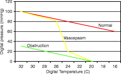
Fig. 29.3
The normal response to cold is a linear decline in digital artery pressure. The vasospastic Raynaud’s patients have a steeper decline in pressure until a threshold temperature is reached, then near instantaneous occlusion. The obstructive Raynaud’s patients have lower normothermic digital artery pressures and a near normal vasoconstrictive response to cold
Noninvasive Diagnostic Techniques
Although the physical examination is frequently completely normal in patients with Raynaud’s syndrome, all noninvasive vascular laboratory testing of the upper extremities should be preceded by a physical examination. Obviously, a complete pulse examination should be performed with attention to the strength and quality of pulses as well as the presence of aneurysms or bruits. In addition, the fingers should be carefully inspected for ulcers. Hyperkeratotic areas may suggest healed ulcers. Hands and fingers should be examined for telangiectasias, skin thinning, tightening, or sclerodactyly, findings suggesting autoimmune disease. Signs and symptoms of nerve compression syndromes should also be sought. Carpal tunnel syndrome is seen in about 15% of Raynaud’s patients [5].
Segmental Arm Pressures
Upper extremity segmental pressures are obtained by measuring blood pressure with pneumatic cuffs above the elbow, below the elbow, and above the wrist while insonating the radial or ulnar artery at the wrist using a continuous wave Doppler. Doppler-derived or plethysmographic waveforms are also recorded at the different levels. Abnormal waveforms or pressures indicate arterial disease proximal to the site where the waveform or pressure was obtained.
A systolic pressure measurement is taken from the upper arm (brachial artery) and at the wrist (radial and ulnar arteries). A 12-cm blood pressure cuff is usually sufficient for measuring the brachial artery pressure. A 10-cm blood pressure cuff just above the wrist is used to measure radial and ulnar artery systolic pressures. Normally, there is no significant systolic pressure gradient between the brachial and wrist levels. A normal wrist/brachial blood pressure ratio is therefore 1.0. A blood pressure difference between the two arms of more than 15 mmHg is indicative of a stenosis or occlusion on the side of the lower pressure. Decreased pressures and/or abnormal waveforms at the above-elbow cuff site indicate occlusive disease at or proximal to the elbow. Normal above-elbow pressures with abnormalities at the above-wrist sites indicate brachial and/or proximal ulnar/radial arterial occlusive disease, respectively (Fig. 29.4). A blood pressure difference between levels or between the radial and ulnar arteries of more than 15 mmHg also indicates an occlusive lesion.
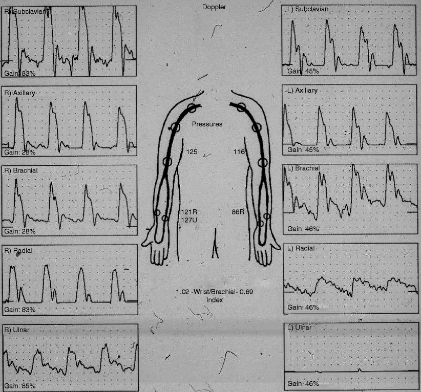
Fig. 29.4
Segmental arm pressures and waveforms demonstrating abnormal pressures and waveforms at the radial and ulnar arteries in the left upper extremity. The study indicates arterial occlusive disease distal to the brachial artery in the left upper extremity
Digital Pressures and Plethysmography
Digital pressure measurements and digital plethysmography are extremely useful in the evaluation of upper extremity ischemia [6]. Photoplethysmography (PPG) or strain gauge plethysmography are both used to measure digital blood pressure and to obtain pulse waveforms. PPG is preferred by most vascular laboratories because the equipment is easier to use and more durable. PPG also allows recording of volume pulses from the tips of the fingers. This can help to document obstruction within digital arteries themselves.
To measure digital pressures, the PPG photo cell is attached to the fingertip pulp with double-sided tape or small strain gauges are placed around the fingertip. One-inch (2.5 cm) blood pressure cuffs are placed around the proximal phalanx. Waveforms are recorded using pulse tracings obtained at high speed to facilitate evaluation of the shape of the waveform. Upstroke time of the waveform should be <0.2 s. Normal waveforms may or may not have a dicrotic notch (Fig. 29.5a). An obstructive waveform has a rounded peak with an upstroke time that is prolonged (Fig. 29.5b). Finger PPG waveforms are not quantitative. Waveform amplitude is therefore not important with amplitude dependent on the gain setting, not blood flow.
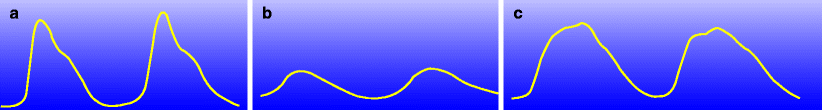
Fig. 29.5
(a) Schematic of a normal PPG waveform with rapid upstroke and typical dicrotic notch. (b) Schematic of an obstructive PPG waveform with prolonged upstroke and rounded peak. (c) Peaked pulse PPG waveform. The dicrotic notch is shifted up on the waveform
Patients with vasospasm will often have an abnormally shaped waveform termed a “peaked pulse.” This is thought to represent abnormal elasticity and rebound of palm and digital arteries (Fig. 29.5c).
Finger blood pressures are measured by inflating cuffs placed at the base of the fingers. Pulsations are recorded at reduced chart recorder speeds as the cuffs are inflated. After digital pulsations are obliterated, the cuff is slowly deflated until the pulsation returns. This pressure is the digital artery pressure.
Stay updated, free articles. Join our Telegram channel

Full access? Get Clinical Tree


