Niche Devices: Atherectomy, Cutting Balloon, and Laser


Niche Devices: Atherectomy, Cutting Balloon, and Laser


Robert J. Applegate MD, FACC, FAHA, FSCAI


ATHERECTOMY


Device

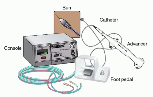
The rotablator (Boston Scientific, Natick, MA) is an over-the-wire system that consists of a nickel-plated diamond-coated brass burr attached to a drive shaft, which can achieve speeds up to 200,000 rpm driven by compressed gas (Fig. 13-9). The 20- to 30-µm-sized diamond chips are located only on the front half of the olive-shaped burr. Much like a high-speed sander, the rotablator burr ablates and creates microparticulate debris when the burr comes into contact with relatively inelastic tissue. The turbine unit is cooled using a saline flush solution, which also helps irrigate the vessel during activation of the burr, helping disperse the microparticulate debris through the vasculature. The reusable console can be adjusted to achieve burr speeds up to 200,000 rpm in the fully activated mode, and speeds of 80,000 rpm when used in the Dynaglide mode (used almost solely for removing the burr from the guide catheter). Each burr and drive shaft comes as a separate unit and is attached to a disposable advancer using a locking mechanism. The advancer has a control knob that allows the operator to advance or retract the spinning burr, and has a range of 10 cm before the burr position needs to be moved if additional atherectomy is desired. The burr and drive shaft accommodate a 0.09″ guide wire with a floppy tip of 30 cm, which has a 0.21″ olive at the joint between the flexible tip and the shaft of the guidewire to prevent the burr from advancing to the flexible tip of the guidewire. A special wire clip is attached at the end of the guidewire as it exits the advancer, and helps prevent the wire from spinning during the high-speed rotation. The advancer also has an internal brake, which prevents the wire from spinning or advancing, which can be manually overridden by a “brake defeat” button at the back end of the device.


FIGURE 13-9 Composite images of the components of the rotablator system. The 0.009″ guidewire used to deliver the burr to the lesion site is not shown.


Principles

The atheroablative effect of the rotablator system is based on the concept of differential cutting, that is, selective ablation of relatively inelastic materials such as calcified or heavily fibrotic atheromatous plaque versus sparing of elastic nondiseased vessel segments. The analogy of rotablation is shaving with the razor, preferentially cutting the hair (inelastic tissue) and not the skin (elastic tissue). The microparticulate debris generated during atherectomy ranges from <5 to <12 µm, depending on the atherectomy speed and composition of the plaque. The debris passes through the coronary microcirculation and is ultimately taken up by the reticuloendothelial system of the spleen and liver. The rate and volume of microparticulate debris in relation to coronary flow will ultimately determine whether or not microvascular obstruction occurs, overwhelming the capacity of the capillary system. The rotablator device also takes advantage of orthogonal displacement of friction as a result of the high rotational speeds, which essentially eliminates the longitudinal friction component of resistance. This characteristic distinguishes it from simple balloon and stent catheters’ passive movement within the coronary arterial system and guide catheter.

The optimal speed during an atherectomy procedure has undergone extensive evaluation (1). Extremely low speeds potentially generate larger microparticulate debris and are inefficient in ablation, whereas very high speeds are associated with significant local rises in temperature and potentially thermal-mediated vascular changes, including a propensity for flow reduction. Rates of 140,000 to 160,000 appear to provide an optimal compromise between the efficiency and the extent of atherectomy in relation to local thermal effects and are the currently recommended range of speed for most atherectomy procedures.


Procedure


Burr Selection

The STRATAS trial evaluated whether more aggressive atherectomy using a target of 0.7 to 0.9 burr and artery ratio reduced the rates of restenosis compared with that using a target of 0.6 to 0.8 burr to artery ratio. Restenosis rates were similar and there was a trend toward more complications with the more aggressive strategy (STRATAS)(2). With the advent and success of intracoronary stents, rotational atherectomy has been used almost exclusively for lesion preparation prior to stenting, when a lesion is undilatable or extensive calcification is present. In contemporary use, burr sizes of 1.25 to 1.75 mm generally provide appropriate lesion preparation prior to stenting, and only under unusual circumstances, burr sizes beyond this would be chosen. In general, the greater the vessel angulation and/or extent of calcification, the smaller the burr sizes that would be chosen. The operator’s comfort level will also help dictate whether the procedure will start with a 1.25- or 1.5-mm burr.


Guide Catheter Selection

Guide catheter selection for rotational atherectomy requires coaxial alignment, appropriate sizing to allow advancement and retraction of the burrs, and some measure of coronary flow during the procedure to help move microparticulate debris through the coronary microvasculature. The internal diameter of the guide catheter should be 0.04″ larger than the burr to minimize difficulty during advancement and retraction of the burr. In addition, guide shapes require gentle transition from the shaft of the burr to the tip as acute angles, such as those that may be seen with a typical Judkins catheter, which can impede the advancement and/or retraction of the burrs at the primary and secondary curves of the catheter. Finally, side holes are recommended so that continued flow occurs around the drive shaft of the rotablator device, even during activation and advancement.


Procedural Technique

Atherectomy is begun approximately 1 cm proximal to the target lesion with constant flush through the drive shaft controlled by a foot pedal connected to the console in an on-and-off fashion. Expert consensus opinion is that optimal rotational atherectomy involves slow advancement of the burr in contact with the stenotic plaque for approximately 10 to 15 seconds and withdrawal of the burr from the lesion, allowing coronary flow to occur, followed by resumption of 10 seconds of atherectomy. Total atherectomy runs are recommended to last no more than 30 to 45 seconds to minimize the potential of overwhelming the microvasculature with the microparticulate debris. Atherectomy is continued with multiple runs until the lesion has been successfully crossed, the full extent of the 10-cm range of the advancer has been exhausted, or it appears that continued efforts would be futile with the burr chosen.


Adjunctive Techniques

The composition of the fluid used to cool the turbine of the advancer is often augmented to include nitroglycerin as a vasodilator as well as Rotoglide (Boston Scientific, Natick, MA) as a coronary lubricant. The latter consists of a sterile egg white and olive oil emulsion, which, in animal testing, appeared to minimize heat generation, permitting a higher rate of rotation of the burr, if needed. General expert consensus also recommends liberal use of vasopressors to maintain an adequate perfusion pressure during rotational atherectomy. Practically, this is most easily performed with 100-µg bolus injections of Neo-Synephrine. Additionally, liberal use of nitroglycerin throughout the procedure is recommended to enhance coronary flow and to minimize microparticulate obstruction of the microvasculature. Prophylactic pacemakers have been used by some to counteract the bradycardia, severe at times, that may accompany rotablation. However, some operators use aminophylline and/or atropine to minimize atherectomy-associated bradycardia without use of temporary pacemakers.


Indications and Contraindications for Use

The manufacturer’s and generally accepted clinical indications and contraindications for use of rotational atherectomy are shown in Table 13-5. Operator experience will dictate comfort levels with these parameters. Accepted indications are heavily calcified lesions able to be crossed with the rotablator guidewire, as well as undilatable lesions. Accepted contraindications include severe lesion entry or exit angulation, and angiographically visible thrombus or dissection.


TABLE 13-5 Indications and Contraindications for Rotational Atherectomy


Indications


Contraindications


Single vessel atherosclerotic CADa


Lesion cannot be crossed with guidewirea


Multi-vessel atherosclerotic CADa


Last remaining vessel with compromised LV functiona


Restenotic lesionsa


Saphenous vein graftsa


Native vessel CAD with lesion length <25 mma


Angiographic evidence of thrombusa


Heavily calcified lesions


Angiographic evidence of dissection at lesion sitea


Undilatable lesions


CAD, coronary artery disease; LV, left ventricular.


a per instructions for use.


Outcomes

The studies evaluating the clinical efficacy and safety of rotational atherectomy were performed in the late 1990s (see Fig. 13-9). The STRATAS trial compared the strategies of routine debulking (burr-to-artery ratio of 0.6-0.8 with adjunctive percutaneous coronary angioplasty [PTCA]) versus that of aggressive debulking (burr-to-artery ratio of 0.7-0.9 with no- or low-pressure PTCA), and observed no difference in procedural outcomes or 9-month angiographic restenosis or target lesion revascularization (2). Rotational atherectomy was compared with PTCA in the randomized COBRA and DART trials, comparing clinical outcomes in de novo coronary artery lesions (3, 4). Efficacy endpoints, including rates of restenosis, were similar between the two techniques, with significant higher incidence of no reflow after rotational atherectomy compared with PTCA. In the randomized ERBAC trial comparing PTCA, rotational atherectomy, and laser atherectomy, restenosis was actually higher for rotational atherectomy than for balloon angioplasty, and was associated with higher rates of complication (5). In the ARTIST trial, rotational atherectomy was shown to have higher restenosis and major adverse cardiac events (MACE) for in-stent restenotic lesions compared with PTCA (6). As a result of these studies, rotational atherectomy was shown to be at most noninferior to PTCA with respect to restenosis, and associated with higher rates of adverse cardiac events (Table 13-6).

With the advent of the stent era, continued interest in debulking prior to stent placement led to the evaluation of atherectomy as an adjunct to stenting. In the SPORT trial, patients with moderately to heavily calcified lesions were randomized to either rotational atherectomy followed by stenting or PTCA followed by stenting (7). The trial was stopped early, but the available data were presented, which showed that the primary endpoint of restenosis at 9 months was similar between the two groups, whereas MACE were higher with rotational atherectomy.

As a result of these clinical trials and the 2011 update of percutaneous coronary intervention (PCI) guidelines (Table 13-7),
coronary atherectomy is given a Class IIA recommendation for the preparation of fibrotic or heavily calcified lesions that might not be crossed by a balloon catheter or adequately dilated prior-to-stent implantation (level of evidence: C). Coronary atherectomy was given a Class III indication (harm) in the routine treatment of de novo or in-stent restenosis (level of evidence: A) (8).


TABLE 13-6 Rotational Atherectomy Quick Facts


What Is Known


PTCA is less effective and has higher complications in heavily fibrotic and heavily calcified lesions


PTRA efficiently ablates heavily fibrotic and calcified lesions


What These Studies Add


PTRA use was associated with similar and sometimes higher rates of restenosis than PTCA in de novo (COBRA [3]; DART [4]; ERBAC [5]) and in-stent restenotic lesions (ARTIST [6])


PTRA had similar to higher rates of MACE than PTCA in de novo (COBRA [3]; DART [4]; ERBAC [5]) and in-stent restenotic lesions (ARTIST [6])


PTRA before stenting had higher rates of restenosis and MACE than PTCA before stenting (SPORT [7])


MACE, major adverse cardiac events; PTRA, percutaneous rotational atherectomy; PTCA, percutaneous balloon angioplasty.


Complications

The operator should be aware of several procedural complications that are relatively common with the use of coronary atherectomy. Bradycardia can accompany rotational atherectomy, and can be severe. It most commonly is associated with treatment of the right coronary artery, but can be seen with treatment of both the left anterior descending (LAD) and circumflex coronary arteries. Although some operators favor preprocedural placement of a temporary pacemaker, this is often not required with use of aminophylline and atropine, as mentioned above, and can lead to cardiac tamponade as a result of ventricular perforation. Coronary slow flow or no flow is a well-recognized complication of coronary atherectomy. Multiple preventive strategies have been outlined. Factors that have been implicated in slow flow include excessive burr speeds, prolonged atherectomy, as well as burr “deceleration” during atherectomy. The latter is defined as a decrease in more than 5,000 rpm below the average working atherectomy speed, and is signaled in the change in the frequency generated by the device during atherectomy. Although very uncommon, burr entrapment can occur as the burr deeply engages the vessel and plaque, causing stalling or complete cessation of rotation of the device. Once this occurs, coronary ischemia will develop as a result of flow obstruction. Because the device performs atherectomy in a clockwise fashion, it has been suggested that detachment of the burr from the advancer and counterclockwise manual rotation of the burr can help facilitate removal of the device. In extreme circumstances, it may be necessary to perform emergency surgery with surgical removal of the device.


FIGURE 13-10 Magnified image of an inflated cutting balloon showing the exposed atherotomes fixed to the long axis of the balloon.


Only gold members can continue reading. Log In or Register to continue

Stay updated, free articles. Join our Telegram channel

May 28, 2016 | Posted by in CARDIOLOGY | Comments Off on Niche Devices: Atherectomy, Cutting Balloon, and Laser

Full access? Get Clinical Tree

Get Clinical Tree app for offline access

TABLE 13-7 2011 ACCF/AHA/SCAI PCI Guidelines