Myocardial Preservation
Myocardial protection has clearly made open-heart surgery a safe and reproducible technique. There continue to be many modifications of the chemical composition of the cardioplegic solution, the optimal temperature (cold or warm), and the route of infusion (antegrade or retrograde). As the concepts of myocardial preservation and surgical approaches have evolved, improved cannulas and cardioplegia delivery systems have been introduced.
Aortic Root Infusion Technique
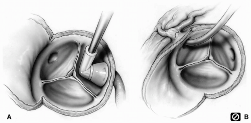
The cannula is introduced into the root of the aorta through a 4-0 Prolene, one and a half circle purse-string suture that is snugged down and secured to the cannula. Although any large-bore needle or cannula is satisfactory, those with a trocar introducer and a side arm for direct intraaortic pressure monitoring are most useful. The side arm can also be used for venting.
Distortion of, or insufficient pressure in, the aortic root may prevent adequate coaptation of the aortic valve leaflets, as will aortic valve insufficiency. The cardioplegic solution passes through the open valve and overdistends the left ventricle, which can cause direct myocardial injury. Digital pressure on the right ventricular outflow tract at the level of the aortic annulus may produce coaptation of the leaflets and prevent regurgitation of the cardioplegic solution.
Excessive infusion pressure can traumatize the coronary arteries, resulting in ischemic myocardial injury. Accurate monitoring of the infusion pressure in the aortic root can be satisfactorily accomplished from the side arm of specially designed cannulas.
Air embolism to the coronary arteries can cause serious myocardial injury. Every effort must be made to clear the cardioplegic line of any air bubbles. A bubble trap is now incorporated into cardioplegia administration systems to minimize this possibility.
Impurities and particulate matter may be present in the cardioplegic solution and can occlude terminal coronary arteries, causing myocardial injury. Quality control in the preparation of the cardioplegic solution prevents such complications.
Between infusions, the cardioplegic solution remaining in the tubing warms up. The warm solution should be flushed out through the free arm of the Y connecting tube before the next infusion into the coronary system.
Uniform cooling of the myocardium by infusion of cold cardioplegic solution is an integral part of myocardial protection. Temperature probes in various parts of the septum and ventricular wall may be used to monitor myocardial temperature during the course of the surgery.
Despite all precautions to keep the heart cool, the anterior surface of the heart tends to rewarm because of the ambient air temperature and the heat radiated from the operating room lights. A gauze pad soaked with cold saline and ice placed over the heart provides additional protection for the right ventricle.
Placement of an insulating pad, a commercially available cooling “jacket”, or a cold lap pad behind the heart can minimize rewarming of the heart by the warmer blood in the descending aorta during the cardioplegic arrest. Care must be taken to avoid cold injury to the left phrenic nerve.


