Fig. 12.1
Resolution of MAT. Fifty percent of patients (indicated by broken line) were free of arrhythmia 5 months after diagnosis. The dotted line indicates a 95 % confidence interval. [Reprinted from Bradley DJ, et al. The clinical course of multifocal atrial tachycardia in infants and children. J Am Coll Cardiol 2001;38(2): 401–408. With permission from Elsevier.]
Electrocardiographic Features
MAT is typically very irregular and can be rapid, with atrial rates as high as 400 beats per minute (Fig. 12.2). Care must be taken to identify different P-wave forms on the ECG, as the isoelectric period between discrete p-waves distinguishes this rhythm from the other irregularly irregular rhythms such as atrial fibrillation or atrial flutter with variable ventricular conduction. Often readily apparent on a surface ECG, the use of adenosine, beta-blockers, and calcium channel blockers has been described to slow the ventricular response allowing better delineation of the underlying p-wave morphology.
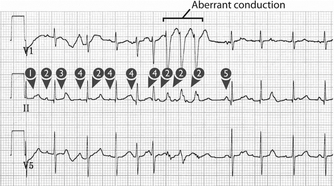
Fig. 12.2
Three electrocardiograph leads (V1, II, V5) in a 3-month-old boy with MAT. Note the five different P-wave morphologies (arrows), the fifth being that of sinus activation. Note also the aberrant conduction (complexes 8–10 from the left). [Reprinted from Bradley DJ, et al. The clinical course of multifocal atrial tachycardia in infants and children. J Am Coll Cardiol 2001;38(2): 401–408. With permission from Elsevier.]
The effort to find the requisite P-waves for the diagnosis is often prompted by MAT’s signature ECG appearance, which includes scattered, aberrantly conducted beats and pauses, due to sinus node suppression after a run of MAT beats as well as blocked conduction of premature atrial impulses (Fig. 12.2). Extended recordings demonstrate periods of sinus rhythm alternating with MAT in most patients.
Mechanism
Just as there is some disagreement about the correct term for this arrhythmia (some authors prefer the term “chaotic atrial tachycardia”), a conclusive mechanistic explanation for MAT remains elusive. Abnormal automaticity, ectopic foci, and triggered activity have all been suggested. Proponents of the term “multifocal” infer that different P-wave forms and irregular PP and PR intervals imply the presence of several ectopic foci in the atria. Ectopic atrial tachycardia, where a single ectopic focus is the source of the tachycardia, exhibits many of characteristics of MAT including resistance to cardioversion and a “warm up” and “cool down” response. However, if an ensemble of ectopic foci gives rise to the arrhythmia, it is unexpected that they would trigger each other’s depolarization, rather than each creating spontaneous, unifocal atrial runs at separate times.
< div class='tao-gold-member'>
Only gold members can continue reading. Log In or Register to continue
Stay updated, free articles. Join our Telegram channel

Full access? Get Clinical Tree


