Fig. 9.1
Auscultation Findings of Patient Case. This figure illustrates auscultation findings of a 85-year-old male who presented with chest tightness and new systolic murmur. It demonstrates the classic findings of a mitral regurgitation murmur: holosystolic murmur with radiation towards the axilla along with a S3 heart sound. Other findings of mitral regurgitation include left atrial enlargement, left ventricular hypertrophy pulmonary hypertension
His arterial pulses demonstrated a brief rapid upstroke.
Test Results
Electrocardiogram demonstrated atrial fibrillation, septal myocardial infarction, right bundle branch block, left anterior fascicular block, left ventricular hypertrophy, and primary T wave changes (Fig. 9.2).
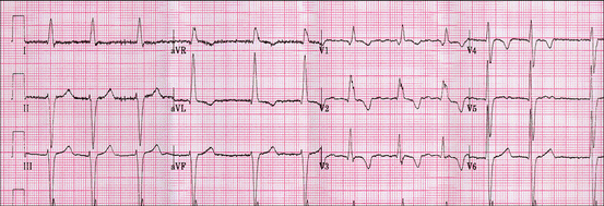
Fig. 9.2
This presents the patient’s previous ECG result obtained in 2004. The ECG shows atrial fibrillation (no true P waves and ventricular rate is irregular), right bundle branch block (wide QRS complexes with terminal R wave in lead V1 and slurred S wave in lead V6), along with septal infarction, left ventricular hypertrophy, and 1° T wave changes
Echocardiography revealed a decreased left ventricular ejection fraction of 40–45 % and left ventricular hypertrophy with a left ventricular internal diastolic dimension of 62 mm. Other findings included: left atrial enlargement, AV sclerosis, 3+ mitral regurgitation (MR), centrally directed, through a morphologically normal mitral valve (MV), and moderate tricuspid regurgitation at 2.6 m/s.
Clinical Basics
Normal Anatomy (Fig. 9.3)
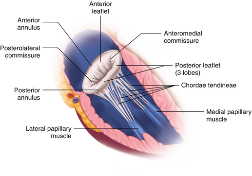
Fig. 9.3
Internal structures of the left ventricle, showing the anterior view of the left ventricle with normal mitral valve anatomy
The mitral valve is a bicuspid valve that separates the left atrium (LA) and left ventricle (LV). The fibrous mitral annulus sits at the left atrio-ventricular junction and serves as an insertion point for the anterior and posterior leaflets.
The valve leaflets open into the left ventricle, tethered by papillary muscles on the wall of the LV that extend chordae tendineae attachments to the leaflets. The anterolateral papillary muscle attaches chordae tendineae to the anterior leaflet while the posteromedial papillary muscle attaches chordae tendineae to the posterior leaflet.
During diastole, the mitral valve is open allowing for LV filling. The mitral valve closes during systole due to increased pressure of the LV, blocking blood flow back into the left atrium. Complete mitral valve closure ensures the unidirectional flow of blood from the LV to the aorta.
The mitral valve closure is associated with the S1 sound.
Definitions
Mitral Valve Regurgitation (MR) occurs when the mitral valve becomes incompetent as a result of compromised or structurally disrupted components of the valve apparatus. This renders gaps in leaflet apposition, allowing for insufficiency, or regurgitation, from the left ventricle back into the left atrium during systole.
There are two main types MR: Chronic and Acute.
Chronic MR results from progressive deterioration of the mitral valve apparatus with increasing insufficiency over the course of many years. The defect may be within the valve itself, referred to as organic, or may appear secondary to pathology within the ventricles without coexisting structural change of the valve, called Functional MR [1, 2].
As a result of any defect, increased volumes within the left side of the heart lead to compensatory dilatation of the left atrium and left ventricle. The resulting increase in LV end-diastolic volume allows for an increased ejection fraction (0.5–0.6), while the enlarged atrial cavity better accommodates the regurgitant volume of blood. These compensations abate the symptoms of MR: pulmonary congestion and forward failure.
Over the course of 6–10 years, however, the prolonged burden to volume overload causes LV dysfunction and the emergence of MR symptoms [3].
Acute MR is the result of an acute impairment of the mitral valve apparatus without time to establish sufficient compensatory mechanisms. The left atrium and left ventricle experience a sudden volume overload as a result of dramatic backflow of blood coupled with decreased stroke volume and cardiac output. The regurgitant volume increases intra-atrial pressure, preventing flow of blood from the pulmonary circulation into the left atrium and resulting in pulmonary congestion. In acute MR, therefore, the patient experiences reduced forward output, or even shock, along with congestion of the pulmonary vasculature [3].
Etiology
MR occurs due to a defect to any part of the mitral valve apparatus – the leaflets, chordae tendineae, papillary muscles, or mitral annulus. The location and extent of damage to the apparatus determines whether the pathology presents as Acute or Chronic MR.
Acute MR may be the result of any sudden event that renders one or several component(s) of the valvular apparatus incompetent. Chordae tendineae rupture, as a result of any etiology, is the most common cause of acute MR. Possible etiologies of acute MR include:
Ischemic Rupture of Chordae Tendineae or papillary muscles: This often results from acute myocardial infarction. If ischemic damage is sudden and severe enough, rupture of the papillary muscles or chordae tendineae will likely result in Acute MR.
Infectious endocarditis is most frequently affects mitral valve structures [4]. It may lead to rupture of the chordae tendineae and/or enlargement of the posterior portion of the mitral annulus [1].
Acute or Rapidly developing Cardiomyopathy.
Other acute causes: Trauma, Surgery and spontaneous rupture.
Chronic MR results from slow, progressive deterioration of the mitral valve apparatus leading to increasingly severe regurgitation across the mitral valve. Common etiologies of Chronic MR include:
Myocardial infarction/Coronary Artery Disease– Prolonged ischemia and/or infarction to the region of the tensor apparatus weakens the papillary muscles and results in an inability to sufficiently tether the corresponding leaflet to the left ventricle. An alternate hypothesis suggests that an infarct near the base of the papillary muscle may cause disorganized muscle contraction, shifting the position of the leaflet during ventricular contraction and precluding appropriate closure [5]. Others argue that MR results from displacement of papillary muscles as a result of left ventricular remodeling rather than an intrinsic defect with the muscle itself [1]. Regardless of the exact mechanism, MR in the setting of ischemic disease is rarely organic or acute, but is rather Functional MR as a result of left ventricular or papillary muscle pathology [1]. Note: Occlusion in the right coronary artery will affect the posteromedial structures, whereas left anterior descending or circumflex artery occlusion will compromise the anteromedial structures. Due to its single blood supply, posteromedial rupture is more common.
Myxomatous degeneration of mitral leaflet(s): Pathological weakening of the valve connective tissues renders the mitral leaflets floppy and redundant, resulting in mitral valve insufficiency. Degenerative MR is usually associated with mitral valve prolapse. Myxomatous degeneration is the most frequent cause of surgical MR [4].
Rheumatic Fever: Acquired during childhood, Rheumatic Fever (RF) induces a slow progression of scarring and retraction of the valvular leaflets that manifests as MR and mitral stenosis with fusion of the commissures in middle age. These cardiac sequelae are no longer common in the United States, but are still a frequent cause of MR in countries where RF remains endemic [1].
Dilated Cardiomyopathy: Dilatation of the LV expands the mitral annulus causing incompetence of the valvular apparatus.
Mitral annular calcification: More common in elderly, mitral annular calcification is usually of little clinical significance; however, it may result in MR due to impairment of contraction of the valve annulus and deformation of valve leaflets. Often associated with conditions such as hypertension, aortic stenosis, diabetes and end stage renal disease [4].
Other less common causes include congenital connective tissue disorders (such as Marfan Syndrome, Ehlers-Danlos Syndrome and Osteogenesis Imperfecta), Hypertrophic Cardiomyopathy, Anorectic Drugs (Fenfluramine and Desfenfluramine), Anthracycline Chemotherapy, Radiation Injury, Carcinoid heart disease, and Congenital parachute valve.
Signs and Symptoms
Depending on the cause, time course of valvular degeneration/destruction, and severity of valvular pathology, MR may remain asymptomatic or lead to severely debilitating symptoms.
Chronic MR (more common) – While compensatory mechanisms are initially successful at maintaining LVEF, continuing pathology leads to symptoms of heart failure and pulmonary hypertension. Of note, patients with an asymptomatic chronic mitral regurgitation may become symptomatic after the imposition of a primary hemodynamic stressor such as pregnancy or infection.
Symptoms of chronic MR include:
Progressive dyspnea upon exertion.
Increasing orthopnea from elevated pulmonary pressures.
Fatigue.
Palpitations from LA dilation leading to arrhythmias (e.g., atrial fibrillation).
Acute MR – Symptoms of acute MR are markedly more severe than those of chronic MR, as the LA and LV have not been able to adapt to the regurgitant blood flow. An immediate and unabated increase in pressure within the LA and LV coupled with a dramatic reduction in cardiac output results in rapid onset of the following symptoms:
Severe dyspnea/orthopnea.
Pulmonary edema or congestion/ pulmonary hypertension.
Weakness.
Anxiety / confusion.
Fever, if stemming from endocarditis.
Prevalence
MR is among the most common valve disease in developed countries, representing nearly one-third of acquired left-sided valve pathology [2].
Moderate to severe mitral regurgitation affects 1.7 % of the general population with equal gender distribution and increases in prevalence with age, from .1 % among 45–50 year-olds to 9.3 % in those ≥75 years [6].
The Framingham Heart Study demonstrates that, including mild MR, this condition is found in 19.0 % and 19.1 % in men and women, respectively [7].
Key Auscultation Features
Chronic Mitral Regurgitation
The classical holosystolic murmur, commencing immediately after S1 and continuing up to S2. It is most often “blowing” and high pitched in quality.
Murmur is heard best over the apex.
The likelihood of auscultating a murmur is proportionately related to the grade of severity of MR on echocardogram. Murmur prevalence increases from 40 % to nearly 90 % with increasing severity of MR to grade 1 through grade 4, respectively [8]. See Figs. 9.4 and 9.5.
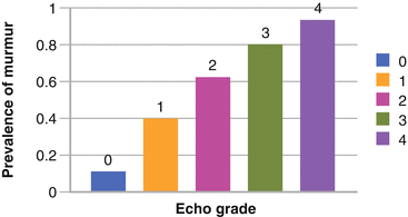
Fig. 9.4
Prevalence of systolic murmurs based upon echocardiographic grade of mitral regurgitation. Note that although mild MR may go undetected by physical exam, most patients with moderately-severe and severe MR have a murmur present (Used with permission from Rahko [8])
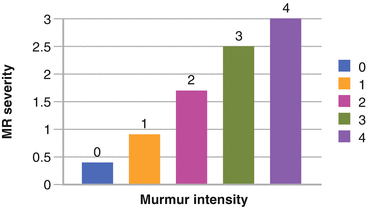
Fig. 9.5
Relationship between murmur intensity and severity of mitral regurgitation. Note that a general relationship exists with more intense murmurs being more likely in the presence of severe mitral regurgitation (Used with permission from Rahko [8])
A widening of S2 may be due to (1) an early A2 with normal P2 indicating a shortened LV ejection time due to an early closure of the aortic valve, or (2) an early A2 combined with delayed P2 indicating the presence of pulmonary hypertension causes the delayed closure of the pulmonic valve.
Presence of S3 gallop.
No S4 gallop heard in chronic MR, due to an enlarged, poorly contractile left atrium.
Auscultation Examples of Mitral Regurgitation.
Click here to listen to an example of an MR murmur in the setting of presumptive rheumatic fever and see an image of the phonocardiogram (Video 9.1).
Click here to listen to an example of moderate to severe MR, as described by Dr. W. Proctor Harvey (Video 9.2).
Click here to listen to an example of a female patient with chronic significant MR, as described by Dr. W. Proctor Harvey (Video 9.3).
Acute Mitral Regurgitation (Fig. 9.6)
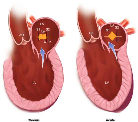
Fig. 9.6
Acute and chronic mitral regurgitation diagrammatic representation (Modified with permission and courtesy of W. Proctor Harvey, MD, MACC, Jules Bedynek, MD and David Canfield. Originals published by Laennec Publishing Inc., Fairfield, NJ, and copyrighted by Laennec Publishing, Inc. All rights reserved.)
An apical systolic murmur with early- and mid- systolic crescendo-decrescendo. The diminishment of the late systolic sound is thought to be due to building atrial pressures that soon equalize that of the LV [9, 10].
Murmur best heard along the left sternal border.
Prominent S4 gallop: Caused by normal-sized, vigorously contracting left atrium following exaggerated expansion during systole. It is suggestive of acute onset of regurgitation due to the rupture of the chordae tendineae that anchor the valvular leaflets [11].< div class='tao-gold-member'>Only gold members can continue reading. Log In or Register to continue
Stay updated, free articles. Join our Telegram channel

Full access? Get Clinical Tree


