Management of Foreign Bodies of the Airway
Julie L. Wei
Lauren D. Holinger
Foreign body aspiration is a serious and potentially fatal occurrence. Through advances in prevention, first aid, and endoscopic technology, a nearly 20% decrease in deaths from foreign body aspiration has occurred within the United States over the last decade. Nevertheless, in 1997, the National Safety Council reported an average of more than eight deaths per day from foreign body aspiration in the United States. Generally, the longer a foreign body has been lodged within the tracheobronchial tree, the greater the morbidity. Thus early diagnosis remains a key to successful and uncomplicated management of these accidents. Although this chapter deals exclusively with children, the same principles apply to the adult population.24
The first extraction of an airway foreign body was performed by Gustav Killian near the end of the nineteenth century, as he describes in the following passage:
On March 27, 1897, whilst eating some soup, [J.W.] aspirated a bone. This accident was followed by attacks of violent cough and dyspnoea, which, however, became gradually less …. On direct laryngeal examination by means of Kirstein’s spatula, the patient being seated with his head strongly deflected to the left, I saw in the right principal bronchus a white mass. On the following day I introduced, under cocaine anaesthesia, a straight tube of 9 millimetres diameter and 25 centimetres length through the larynx and the trachea until I came near the foreign body. The curvature of the trachea was thus removed, and the foreign body could be seen distinctly. I had great difficulty in catching hold of the foreign body, using a pair of slender forceps which had specially and quickly been made. The difficulties were great, as at that time … I was still without the necessary practice which enables one to look easily, and even more to operate, through long tubes. Eventually I succeeded in catching the bone and in extracting it. The patient was able to return home on the following day.20
The basic principles of extraction were meticulously developed by Chevalier Jackson during the first half of the twentieth century. Jackson’s concepts of the various mechanical problems and demonstrations of their solutions remain valid today. At any given institution, airway foreign body problems should be managed by the individuals with the best training and most experience, regardless of their particular subspecialty. As Jackson16 stated and Hughes and colleagues14 reiterated, the techniques of foreign body extraction should be mastered through practice on lung models and then on anesthetized laboratory animals before they are attempted in human beings.
Epidemiology and Etiology
Older infants and toddlers constitute the vast majority of patients with foreign body aspiration. Darrow and Holinger8 reviewed multiple case series and found that children <5 years of age account for approximately 84% of cases and children <3 years account for 73%. The high incidence in this age group reflects the tendency of children to explore the world using their mouths. In addition, these children have not developed a full posterior dentition and may have immature neuromuscular mechanisms for swallowing and airway protection. Moreover, many youngsters are allowed to talk, run, or play with food or other objects in their mouths. For uncertain reasons, boys aspirate foreign bodies more frequently than girls by a ratio of approximately 2:1. In adults, other factors play a role in foreign body aspiration, such as neurologic dysfunction, dental trauma, and aspiration of larger than normal pieces of food, usually associated with alcohol consumption.
Although adults most commonly aspirate bones from fish, birds, or small mammals, children usually aspirate vegetable matter. Darrow and Holinger8 found that nuts, particularly peanuts, account for approximately 34% of cases of foreign bodies found in the pediatric airway. Even nut fragments found in crunchy peanut butter have been aspirated. Other commonly aspirated types of vegetable matter include pieces of raw carrot, apple, dried beans, popcorn, and sunflower, watermelon, or pumpkin seeds. Darrow and Holinger8 found that the most commonly aspirated inorganic objects in school-aged children include tacks, nails, pencil erasers, pins, plastic beads, and even marbles.
The spectrum of airway foreign bodies varies from country to country, depending on the diet and customs of the population. For example, Mu and colleagues23 reported that in mainland China, nearly 95% of aspirated foreign bodies in children were organic. In contrast, more industrialized countries have a greater incidence of aspiration of plastic foreign bodies, because plastic parts are used frequently by the toy industry. Other reported examples of the cultural influence on these accidents include aspiration of Mardi Gras beads, pieces of crab shell, holiday decorations and gifts, and straight pins by Middle Eastern women and girls, who hold the pins between their lips while securing their head scarves. Fortunately, ingestion of safety pins
has become rare since the advent of disposable diapers. Rarely, medical therapy itself may be responsible. Aspiration of pills, a thermometer fragment, and an object within a metered-dose inhaler has been reported.
has become rare since the advent of disposable diapers. Rarely, medical therapy itself may be responsible. Aspiration of pills, a thermometer fragment, and an object within a metered-dose inhaler has been reported.
History
Three stages of symptoms result from the aspiration of an object into the airway:
Initial event. Violent paroxysms of coughing, choking, and gagging, and possibly airway obstruction occur immediately after a foreign body is aspirated. An esophageal foreign body large enough to cause posterior tracheal compression may cause similar symptoms. Such a history can be elicited in most cases, but unfortunately many parents tend to downplay the significance of such an event or do not recall the incident until after the foreign body has been extracted. Some parents engage in wishful thinking and minimize the symptoms, hoping that nothing is wrong and that no surgical intervention will be required. Older children are often reluctant to admit to such an episode for fear of being punished.
Asymptomatic interval. During the second stage, the foreign body becomes lodged, reflexes fatigue, and the immediate irritating symptoms subside. This stage is the most treacherous and accounts for a large percentage of delayed diagnoses and overlooked foreign bodies. It is during this second stage that the physician is inclined to minimize the possibility of a foreign body accident, being reassured by the absence of signs and symptoms.
Complications. In the third stage, obstruction, erosion, or infection develop and again direct attention to the presence of a foreign body. Signs include fever, cough, and hemoptysis. Complications include formation of bronchial granulation tissue, atelectasis, pneumonia, lung abscess, and eventually bronchocutaneous fistula. Such complications occur more rapidly after aspiration of vegetable matter and sharp objects than of plastics and other biologically inert materials.
The current medical practice of treating an asthmatic or “croupy” child with antibiotics or corticosteroids may obscure signs and symptoms that normally would be expected with a retained foreign object. Clearing of symptoms with these agents cannot always be assumed to be diagnostic of a specific disease process. The fact that a wheeze disappears or a pneumonic process temporarily clears may merely mean that the patient’s reaction to a foreign body has been controlled temporarily. The recurrence of “asthma” after tapering of therapy should heighten a physician’s suspicion of an aspirated foreign body.
A positive history must never be ignored. A negative history may be misleading. Choking or coughing episodes accompanied by wheezing are highly suggestive of foreign body aspiration. The literature reveals that diagnosis is delayed >24 hours in 50% of cases, as noted by Wiseman,33 and more than 1 week in 15% of cases, as reported by Reilly and colleagues.27 Disregarding a child’s story because of age or lack of symptoms may cause a delay in diagnosis, which may make removal more difficult and complicated. As Wetmore31 stated, once an aspirated foreign body is suspected, the burden of proof is on the bron- choscopist. Furthermore, Mantor and colleagues21 have suggested that “some negative bronchoscopies are necessary in order to prevent the morbidity that occurs from a missed foreign body aspiration.”
Physical Examination
Laryngeal Foreign Bodies
Large globular foreign bodies that become lodged between the vocal cords usually cause complete obstruction and asphyxiation unless promptly expelled. Flat, thin, and sharp objects, such as eggshell and bone, may become lodged between the vocal cords in the sagittal plane. Dysphonia, croupy cough, stridor, and varying degrees of dyspnea ensue, all of which increase as edema and inflammation progress. Odynophagia may also occur.
Tracheal Foreign Bodies
Jackson and Jackson18 described three features of tracheal foreign bodies: The audible slap and the palpatory thud result from the impact of a mobile foreign body against the tracheal wall on deep inspiration or coughing. The asthmatoid wheeze results from partial bronchial obstruction from the foreign body and the inflammatory reaction. Biphasic stridor may also occur if the foreign body is within the extrathoracic trachea.
Bronchial Foreign Bodies
Wiseman33 noted that the classic triad of wheezing, coughing, and decreased air entry to the obstructed side was present in only 31% of children examined within 24 hours of bronchial foreign body aspiration and only 47% of children examined after 24 hours. Moreover, physical signs may change rapidly with migration of the foreign body and with the development of edema and infection. If the foreign body eventually lodges within one bronchus, the physical examination may stabilize.
Radiologic Evaluation
Inspiratory and expiratory posteroanterior and lateral chest radiography is standard when foreign body aspiration is suspected. If the foreign body is radiopaque, a film is taken in the greatest diameter of the object for accurate localization before endoscopy. More than 90% of foreign bodies, however, are radiolucent, as reported by Vane and colleagues.31 This percentage is likely to increase as more polyethylenes are used to make toys.
Jackson and Jackson19 described the pathophysiology behind the radiographic diagnosis of radiolucent bronchial foreign bodies. Initially, the object creates a bypass valve, which still allows ingress and egress of air. At this stage, radiography results are normal. As edema of the surrounding bronchial wall develops, a check valve is created. On inspiration, the bronchus dilates and permits ingress of air. However, on expiration, the bronchus constricts, and contact of the edematous bronchus with the foreign body blocks the egress of air. Thus, air trapping (obstructive emphysema) results. Radiographically, when a check valve is created, the inspiratory film is normal, whereas the expiratory film shows hyperinflation of the affected lung and shift of the
mediastinum to the opposite side (Fig. 76-1). If inspiratory and expiratory films are not possible because a child is tachypneic or uncooperative, lateral decubitus chest films or fluoroscopy may also identify air trapping. Eventually, when enough edema develops to block both ingress and egress of air, a stop valve is created. Obstructive atelectasis is seen radiographically. This late complication usually takes days or weeks to develop.
mediastinum to the opposite side (Fig. 76-1). If inspiratory and expiratory films are not possible because a child is tachypneic or uncooperative, lateral decubitus chest films or fluoroscopy may also identify air trapping. Eventually, when enough edema develops to block both ingress and egress of air, a stop valve is created. Obstructive atelectasis is seen radiographically. This late complication usually takes days or weeks to develop.
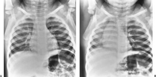 Figure 76-1. A: Anteroposterior radiograph of the chest during inspiration. B: Anteroposterior radiograph of the chest during expiration. Note the trapping of air in the left lung field, caused by a peanut in the left mainstem bronchus. |
Although reports vary, Black and colleagues2 found that inspiratory and expiratory chest radiography was diagnostic in 83% of 440 children with tracheobronchial foreign bodies. In cases of suspected tracheal foreign bodies, posteroanterior and lateral soft tissue neck films (high-kilovoltage airway films) are the radiographic tests of choice. As Esclamado and Richardson9 reported, airway films are abnormal in 92% of children with tracheal foreign bodies, whereas chest radiography is abnormal in only 58%. Pneumomediastinum is a rare but highly diagnostic finding. It was reported by Burton and colleagues6 in 7% of patients with tracheobronchial foreign bodies. When a radiolucent foreign object’s aspiration is suspected and the airway or chest radiography is negative, one may take a radiograph of a duplicate object if available. If the object appears to be radiopaque, then the absence of finding on the radiograph is reassuring.
In rare cases, when a radiolucent foreign object is lodged too far in the lung periphery for endoscopic management, it may be precisely localized by bronchography, computed tomography, or in some cases magnetic resonance imaging. Imaizumi and colleagues15 noted that because of peanuts’ high lipid content, peanut fragments appear as high-intensity signals on T1-weighted images. Such information would confirm the diagnosis and would assist in localization if thoracotomy and segmentectomy were to become necessary.
Treatment
A bronchial foreign body usually does not constitute an acute emergency unless there is complete obstruction of one main bronchus, causing inadequate oxygenation. Supraglottic, glottic, and tracheal foreign bodies are more likely to cause acute emergent situations, with severe respiratory distress. In general, the treatment of choice is reasonably prompt endoscopic removal under conditions of maximal safety and minimal trauma. Too often, foreign bodies are considered emergencies, leading to hasty, inadequate preoperative planning and poorly prepared, improper attempts at removal. Most patients with foreign bodies who have come to the endoscopic surgeon have already passed the acute phase. When no urgent danger to the patient’s life exists, the problem should be approached with complete and thoughtful consideration of the physiologic and mechanical factors involved.
The endoscopic removal can be scheduled once trained personnel are available, instruments have been checked, and techniques have been tested. However, untoward delay of bronchoscopic removal is potentially harmful because the foreign body may become dislodged from the bronchus and impacted in the larynx, causing asphyxiation. Therefore endoscopy is deferred only until preoperative studies have been obtained and the patient has been prepared for surgery by adequate hydration and emptying of the stomach. Foreign bodies in the larynx or tracheobronchial tree are usually removed on the same day the diagnosis is considered.
Two situations exist, however, in which an airway foreign body does constitute an acute emergency: actual and potential airway obstruction.
Actual or Potential Airway Obstruction
The most serious complication of foreign body aspiration is complete obstruction of the airway. Globular food objects—such as hot dogs, grapes, nuts, and candies—are the most frequent offenders, whereas rubber balloons and other toys are common among nonfood objects. Large or multiple esophageal foreign bodies can also cause airway obstruction by posterior compression. Stephen Bradwell remarked on the matter in 1633: “Of Things that endanger stopping of the breath in swallowing, some are Sharp, and some Blunt. … I have heard of a Child in Woodstreet strangled with a Grape.”4
Stay updated, free articles. Join our Telegram channel

Full access? Get Clinical Tree


