Lysis for Iliofemoral DVT
Omar Al-Nouri
Bernadette Aulivola
In the United States alone, venous thromboembolism (VTE) affects some 900,000 individuals. Acute lower extremity deep venous thrombosis (DVT) has significant associated long-term morbidity. Postthrombotic syndrome (PTS) may occur in 40% to 80% of patients with DVT, leading to significant healthcare costs in its long-term management. Prevention is of paramount importance in reducing the long-term complications that may arise after lower extremity DVT. The basis of preventing DVT stems from Virchow’s initial observations over 150 years ago that the pathogenesis of DVT is related mainly to hypercoagulability, venous stasis, and endothelial damage. Methods of DVT prevention are either mechanical, with the goal of improving venous flow and reducing stasis, or pharmacologic, based on agents that act as anticoagulants.
The traditional mainstay therapy for acute DVT has been anticoagulation with unfractionated heparin (UFH) or low molecular weight heparin (LMWH) with conversion to oral warfarin for a specified period of time. Anticoagulation alone significantly decreases the risk of subsequent pulmonary embolism (PE) and reduces the incidence of symptomatic leg swelling in patients with DVT. Despite the dramatic decrease in subsequent PE development with anticoagulation, significant lower extremity venous valvular dysfunction occurs and leads to the development of PTS. Markel et al. demonstrated that despite recanalization of a high percentage of extremity veins treated with anticoagulation, up to 50% of patients had residual chronic thrombus. This leads to a significant inflammatory response resulting in valvular incompetence and dysfunction. Faulty venous valve function and residual venous obstruction despite anticoagulation leads to ambulatory venous hypertension resulting in PTS.
The development of PTS has a significant impact on healthcare costs and leads to a significant decreased quality of life. PTS leading to recalcitrant edema, pain, color changes, and ulcerations can be very problematic to manage. Given this, aggressive treatment of acute DVT with catheter-directed thrombolysis (CDT) is now recognized as an option in first-line therapy. Not all DVTs require treatment with CDT. Patients who derive the greatest benefit are those with extensive iliofemoral DVT. To a lesser extent femoropopliteal DVT may be treated with CDT, whereas DVT in the popliteal segment alone does not warrant treatment with CDT. CDT is most efficacious if initiated shortly after development of iliofemoral DVT, ideally within 1 week.
All patients with iliofemoral DVT should be evaluated for possible CDT. A thorough history should be obtained to identify those patients with relative or absolute contraindications for CDT. Patients with known malignancy, those who are nonambulatory, and postsurgical patients are likely not optimal candidates for thrombolytic therapy. Other absolute contraindications to thrombolytic therapy include active bleeding disorder, gastrointestinal (GI) bleeding within 10 days, stroke within 6 months, head injury within 3 months, and intracranial or spine surgery within 3 months.
The initial management of patients with iliofemoral DVT is therapeutic anticoagulation. We prefer the use of UFH, as it has a shorter half-life than LMWH, can be turned on and off with ease, and can be reversed if need be. A bolus dose of intravenous heparin is administered at 80 units/kg and subsequently, a continuous infusion of IV heparin is administered at 18 units/kg/hr with the goal of keeping the aPTT between 60 and 90 seconds. Alternatively, LMWH therapy may be used with an initial bolus dose of 1 unit/kg followed by twice-daily injections of 1 unit/kg. In preparation for initiating CDT, a Foley catheter should be placed to monitor for hematuria as a complication of thrombolysis. An intensive care unit (ICU) bed should be arranged for the patient. Baseline coagulation labs should be sent off including a CBC, PT, aPTT, INR, and fibrinogen. The vast majority of thrombolysis cases can be performed under local anesthesia with minimal sedation. General anesthesia is usually unnecessary.
Intervention
Patients should receive a prophylactic dose of antibiotics 30 to 60 minutes before beginning of the procedure. The groins and ipsilateral popliteal fossa are shaved. The patient should be placed on continuous cardiopulmonary monitoring while receiving IV sedation.
Positioning
Patients are positioned in the prone position initially to facilitate access to the ipsilateral popliteal vein. Ipsilateral popliteal vein cannulation is preferred as it provides the most direct access to the thrombus decreasing the length of catheters required and decreasing the risk of embolization. Alternatively, contralateral common femoral vein or internal jugular vein are other access sites that may be considered.
Technique
Catheter-directed Thrombolysis Infusion
Prior to initiation of thrombolysis a full coagulation panel including CBC, INR, aPTT, and fibrinogen is established for baseline monitoring. Tissue plasminogen activator (TPA) is the most widely used thrombolytic agent for venous thrombolysis. Local anesthetic consisting of 1% to 2% lidocaine is infiltrated at the access site. Using ultrasound guidance, the ipsilateral popliteal vein is percutaneously accessed with a micropuncture needle. A 4-Fr micropuncture sheath is inserted over a 0.018-in guidewire. The sheath is upsized to a short 5-Fr sheath. An initial venogram is performed through the sheath to identify the extent of thrombus within the venous system. A 5-Fr angled glidecatheter is then advanced over a 0.035-in angled glidewire (Terumo) from the access site, through the popliteal, femoral, common femoral, external iliac, and common iliac veins into the inferior vena cava (IVC). Patency of the IVC is confirmed with a contrast venogram.
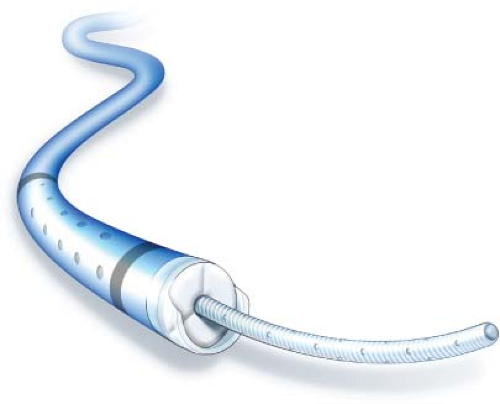 Figure 10.1 Cragg-McNamara valved infusion catheter. |
Multiple microinfusion catheters used to deliver a continuous infusion of thrombolytic agent into the thrombus are commercially available. Our preference is to infuse TPA with the use of a Cragg-McNamara (Fig. 10.1) catheter. This is a single-lumen 5-Fr catheter that is introduced over a wire such that its tip extends into the patent vein just proximal to the thrombus This catheter has a valved end-hole that allows the catheter to be passed over a wire, but when the wire is removed, allows for infusion through side-holes without the need for a tip-occluding wire. In cases of extensive thrombosis, one microinfusion catheter may be inserted through the popliteal vein puncture site for treatment of the femoropopliteal segment, and a second puncture at the popliteal or common femoral vein may be performed for placement of a microinfusion catheter across the iliofemoral segment such that infusion is performed throughout the length of the iliofemoropopliteal system.
Alternatively, the Angiodynamics Pulse Spray (Fig. 10.2) infusion system may be used. This catheter requires the use of a ball-tipped wire that occludes the end-hole of the catheter allowing for infusion of TPA through catheter side-holes. A bolus of 5 to
10 mg of TPA is manually pulsed into the thrombus through the catheter. Continuous TPA infusion is then administered through the microinfusion catheter at a rate of 1 mg/hr. Heparin is infused through the side arm of the sheath at a rate of 500 units/hr. Patients should be closely monitored in an ICU setting with Foley catheter in place to monitor for hematuria. A knee immobilizer should be placed on the ipsilateral lower extremity to prevent kinking or movement of the sheath and catheter. Laboratory values should be checked every 6 hours during CDT therapy, including CBC, INR, aPTT, and fibrinogen. TPA infusion rate should be halved to 0.5 mg hour if the fibrinogen level drops below 150 to 180 mg/dL or decreases more than 50% from baseline.
10 mg of TPA is manually pulsed into the thrombus through the catheter. Continuous TPA infusion is then administered through the microinfusion catheter at a rate of 1 mg/hr. Heparin is infused through the side arm of the sheath at a rate of 500 units/hr. Patients should be closely monitored in an ICU setting with Foley catheter in place to monitor for hematuria. A knee immobilizer should be placed on the ipsilateral lower extremity to prevent kinking or movement of the sheath and catheter. Laboratory values should be checked every 6 hours during CDT therapy, including CBC, INR, aPTT, and fibrinogen. TPA infusion rate should be halved to 0.5 mg hour if the fibrinogen level drops below 150 to 180 mg/dL or decreases more than 50% from baseline.
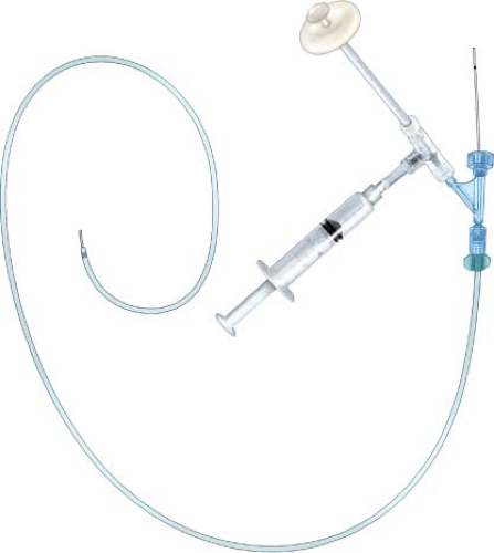 Figure 10.2 Angiodynamics pulse spray infusion system. |
Stay updated, free articles. Join our Telegram channel

Full access? Get Clinical Tree


