Treatment of Superior Vena Cava Syndrome and Central Stenosis
Peter Gloviczki
Sameh Said
Superior vena cava (SVC) syndrome is the collection of signs and symptoms that are observed in patients with obstruction of the venous return to the heart through SVC or any of its major tributaries. Presentations can be acute, developing within a few hours to days or chronic, with progressive appearance of signs and symptoms through several months. The main manifestations include facial or neck swelling, feeling of fullness in the head and neck, dilated neck veins and chest wall collaterals (Fig. 8.1; Table 8.1), arm swelling, shortness of breath, headache, visual symptoms, lightheadedness or syncope, especially when the patient bends over. Patients frequently sleep on several pillows and in severe cases, mental changes are present.
Patients with benign etiology can develop acute thrombosis due to central lines or pacemaker wires, or chronic disease due to mediastinal fibrosis. In acute disease, initial management starts with supportive measures which include supplemental oxygen, elevating the head of the bed and medical therapy which usually includes diuretics, anticoagulation with or without a short course of steroids. Consideration of the primary etiology is important as chemoradiotherapy should be considered in the presence of malignant obstruction, while anticoagulation or catheter-directed thrombolysis plays a role in the presence of a thrombus without a mass compression. Removal of the central line or replacement of the pacemaker wire, if possible, should be considered. In patients with SVC stenosis due to mediastinal fibrosis or obstruction due to chronic thrombosis, percutaneous balloon angioplasty with venous stenting has become the first line of treatment.
Open surgical treatment should be considered for symptomatic patients with benign etiology who are unresponsive to conservative treatment and not amenable to or failed endovascular interventions. SVC reconstruction to treat SVC syndrome caused by malignancy is indicated in those patients who undergo thoracotomy for surgical resection of the tumor.
 Figure 8.1 Dilated chest wall collaterals are one of the characteristic features of superior vena cava obstruction. |
Adequate inflow is required for patients who undergo SVC reconstruction. Either one of the innominate veins or one of the internal jugular veins must be patent to assure long-term success of bypass graft that is placed on the central SVC or the right atrial appendage. Also, the patient should be fit for partial or full median sternotomy. Metastatic disease, not amenable for surgical treatment, is a contraindication for surgical SVC bypass.
Evaluation includes duplex ultrasonography and computed tomographic (CT) or magnetic resonance (MR) venography to establish the benign or malignant cause of the syndrome, to exclude underlying primary or metastatic malignancy, and to establish the extent and acuteness of the obstruction. If medical management fails, endovascular treatment is not possible or was unsuccessful, planning of the operation begins.
The first step in operative planning is to understand the venous anatomy and this usually requires noninvasive imaging of the major intrathoracic veins and the SVC. Ultrasound of the jugular and subclavian veins confirms patency of these veins and helps to identify site of the distal anastomosis of a graft. Axial imaging through a CT or MR venography identifies the etiology of SVC syndrome (Fig. 8.2) and whether it is due to the presence of an extrinsic mass effect or intraluminal thrombus. Preoperative bilateral upper extremity contrast venography still provides the best roadmap needed for surgical treatment (Fig. 8.3). It identifies the level of obstruction and helps in planning the technique of reconstruction or bypass mainly determining the inflow and the outflow of the bypass graft that will be used in surgery.
Table 8.1 Common Manifestations of Superior Vena Cava Syndrome | ||||||||||||||||||||||||
|---|---|---|---|---|---|---|---|---|---|---|---|---|---|---|---|---|---|---|---|---|---|---|---|---|
|
 Figure 8.2 Preoperative computed tomography of the chest is important in identifying the etiology of superior vena cava syndrome: In this patient it was due to fibrosing mediastinitis for an old histoplasmosis. Notice the dense calcifications in the mediastinum close to the superior vena cava (white arrows). |
In the cases, in which an autogenous saphenous or femoral vein will be used, preoperative Duplex scan of lower extremity veins is done to confirm the size, patency of the veins, and rule out previous deep vein thrombosis.
Open surgical reconstruction remains an excellent option in selected patients. Several surgical techniques have been described including patch venoplasty in the presence of central vein stenosis or bypass. Possible bypass graft types include autografts such as saphenous vein graft or femoral vein graft, or spiral vein graft. Externally supported expanded polytetrafluoroethylene (ePTFE) graft is an excellent conduit, rarely homograft, or a graft using autologous or bovine pericardium has been used.
Spiral saphenous vein has been our preferred conduit followed by ePTFE graft. The femoral vein is also an excellent alternative, although some limb swelling, especially in young patients, can be an unintended side effect.
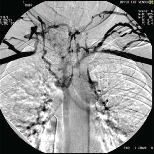 Figure 8.3 Preoperative bilateral upper extremity venography provides an important roadmap before surgery. Notice the multiple collaterals and the complete occlusion of both innominate and jugular veins. |
Positioning
The patient is positioned supine with both arms tucked in. Both sides of the neck and the chest is prepped and draped in sterile fashion. If the plan is to use a saphenous or femoral vein as the bypass graft, one or both lower extremities are prepped and draped down to the knee joint, with the thighs in the frog-leg position.
Intraoperative and Anesthetic Management
Clamping of the SVC in these cases is usually well tolerated because of the preexistent chronic occlusion; on the other hand if the SVC is patent and is required to be clamped for venous reconstruction there is a potential risk of increase in intracranial pressure with subsequent brain edema postoperatively and a decrease in the cardiac output should be anticipated.
It is preferable to have radial arterial and central venous jugular lines inserted in these cases to obtain continuous pressure monitoring although placement of a jugular line should not interfere with surgical plans or clamping. Volume expansion may be required during the period of venous clamping; therefore it is better to have two large peripheral intravenous accesses in the femoral veins.
The following are some intraoperative protective strategies that may be required:
Control of venous bleeding and blood replacement. Median sternotomy in these patients may result in considerable bleeding. Many patients may require rapid surgical control and volume replacement with colloids, blood, and blood products to increase venous return and maintain a normal cerebral arterial–venous gradient. Excess volume can be removed with diuretics postprocedure.
Vasoactive agents to increase the mean arterial blood pressure and maintain cerebral perfusion.
Anticoagulation is mandatory before venous clamping and in the postoperative period.
Surgical Options
Tangential Resection and/or Venoplasty
If the SVC is not totally occluded, partial resection can be performed and the defect is closed either primarily after adequate mobilization of the proximal and distal ends or with the use of a patch. Different patch material can be utilized including autologous or bovine pericardium or a saphenous vein patch.
SVC Bypass or Replacement
This is the most common type of reconstruction and different replacement materials have been described.
Stay updated, free articles. Join our Telegram channel

Full access? Get Clinical Tree


