EVLP donors
Year
DBD
DCD
EVLP → Lung transplant
Ingemansson
Ann Thor Surg
2009
×
Yes
Pego-Fernandez
Rev Bras Cir Cardiovasc
2010
×
No
Cypel
New Engl J Med
2011
×
×
Yes
Madeiros
J Heart Lung Transpl
2011
×
No
Sedaria
Ann Thor Surg
2011
×
No
Cypel
J Thorac Cardiovasc Surg
2012
×
×
Yes
Aigner
Am J Transplantation
2012
×
Yes
Valenza
Transp Proc
2012
×
Yes
Zych
J Heart Lung Transpl
2012
×
×
Yes
Wallinder
J Thorac Cardiovasc Surg
2012
×
Yes
Wallinder
Eur J Card-Thor Surg
2013
×
Yes
29.2 Technique
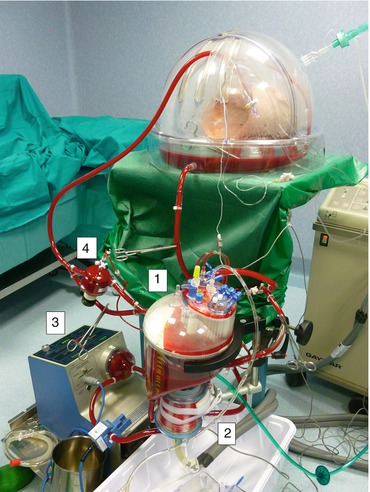
Figure 29.1 shows the circuit used to perfuse the isolated lungs. It consists of a blood reservoir (1 in the figure) connected to a gas oxygenator with a built-in heat exchanger (2), a centrifugal pump (3), a leukocyte arterial filter (4), and a non-heparin-coated polyvinyl tubing. The system is primed with Steen solutionTM (Vitrolife, Gothenburg, Sweden). This is a specifically designed buffered solution with an extracellular-type composition and with an optimized albumin-based colloid osmotic pressure. Methylprednisolone, antibiotics, and heparin are also added to the perfusate. To run the EVLP, the lungs procured from donors and cold stored on ice are contained in a specifically designed chamber (XVIVO, Vitrolife). Temperature of the perfusate is gradually increased to a target temperature of 37 °C over approximately 30 min. Once the lung outflow temperature exceeds 32 °C, mechanical ventilation of the lungs is started. The circuit oxygenator is used unconventionally during EVLP; in fact, gas flow through the artificial lung is composed of CO2 and air and is intended to add CO2 and remove O2 so that the perfusate composition is similar to that of the pulmonary artery. The lungs are ventilated and perfused up to 4 h in most protocols, at the end of which, a final evaluation of lung function is performed. This takes into account parameters of lung perfusion (perfusate flow, temperature, and pulmonary artery pressure, pulmonary vascular resistance) and ventilation (tidal volume, airway pressure, dynamic compliance, respiratory rate, PEEP and FiO2), together with analysis of partial pressures of oxygen (PO2) and carbon dioxide (PCO2). Chest X-ray and fibrobronchoscopy are also added to the final evaluation of lung suitability. If deemed suitable for transplantation, the lungs are flushed with preservation solution and cold stored on ice, ready to be used for transplantation.


Fig. 29.1
Figure shows the circuit used to perfuse the isolated lungs. It consists of a blood reservoir (1) connected to a gas oxygenator with a built-in heat exchanger (2), a centrifugal pump (3), a leukocyte arterial filter (4), and a non-heparin-coated polyvinyl tubing
While EVLP protocols all account for reperfusion, reconditioning, evaluation, and cooling periods, two main philosophies have been diversified over time, summarized in the Toronto and the Lund protocols. The main differences are shown in Table 29.2.
Table 29.2
Comparison between the Lund and Toronto EVLP protocols
Lund | Toronto | |
|---|---|---|
Duration, hours | 1.5 | 4 |
Perfusate flow, % donor CO | 100 | 40 |
Pulmonary artery pressure, mmHg | <20 | 10–15 |
Left atrium | Open | Closed |
FiO2, % | 50 | 21 |
Tidal volume, mL/kg donor’s weight | 5–7 | 7 |
Respiratory rate, bpm | 15–20 | 7 |
Perfusate composition | Cellular | Acellular |
29.3 Clinical Application
Ex vivo lung perfusion technique is used to evaluate and/or recondition the function of lungs procured from marginal donors.
Stay updated, free articles. Join our Telegram channel

Full access? Get Clinical Tree


