and Alessandra Cancellieri2
(1)
Department of Radiology, Bellaria Hospital, Bologna, Italy
(2)
Department of Pathology, Maggiore Hospital, Bologna, Italy
Radiology | Giorgia Dalpiaz |
Pathology | Alessandra Cancellieri |

Dark lung pattern | Definition | Page 234 |
Subset bronchiolar | Page 235 | |
Subset vascular | Page 237 | |
Tables | Page 238 |
Dark Lung Pattern
Definition
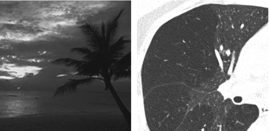
A dark lung pattern is present when variable portions of pulmonary parenchyma show reduced attenuation to the X‐rays, and then are darker than normal. The size and number of the vessels within the pathologic dark areas are smaller than in the non-pathologic white areas.
Unlike the cystic pattern, here the basic abnormality is not pure black, but rather a dark gray. The dark areas basically depend on a reduction of blood flow, either due to a reduced vascular flow (vascular dark lung) or to a hypoxic vasoconstriction from a bronchiolar disease (bronchiolar dark lung).
Mosaic oligoemia/perfusion


The non-pathologic “white” areas are also defined “pseudo-GGO”. When the “mosaic” is otherwise given by patchy areas of ground-glass opacity (GGO), size and number of the vessels within different areas should be equal.
The prevalent distribution of the signs together with the presence or absence air trapping and non-parenchymal signs may be helpful for the diagnosis of a specific disease (see the Tables at the end of this Chapter).
Castañer E (2009) CT diagnosis of chronic pulmonary thromboembolism. Radiographics 29(1):31
Hansell DM (2008) Fleischner society: glossary of terms for thoracic imaging. Radiology 246(3):697
Kligerman SJ (2015) Mosaic attenuation: etiology, methods of differentiation, and pitfalls. Radiographics 35(5):1360
Maffessanti M, Dalpiaz G (2011) Dark lung pattern. In: Leslie KO, Wick MR (eds) Practical pulmonary pathology. A diagnostic approach. Churchill Livingstone (Elsevier), Philadelphia
Subset Bronchiolar
In patients with dark lung pattern from bronchiolar diseases, the dark areas ( ) result as a consequence of bronchiolar narrowing (►) or obstruction (
 ) with subsequent hypoxic vasoconstriction and variable air trapping.
 ) result as a consequence of bronchiolar narrowing (►) or obstruction (
 ) with subsequent hypoxic vasoconstriction and variable air trapping.
Stay updated, free articles. Join our Telegram channel

Full access? Get Clinical Tree


