Intravenous Drug Injection into Femoral Artery
JEFFREY R. RUBIN and YEVGENIY RITS
Presentation
A 63-year-old man underwent noninvasive vascular studies for a pulsatile ulcerated mass in the right groin. The patient had been admitted to the medicine service with complaints of fever and chills with a history of intravenous drug use since the age of 14. The patient admits to a recent injection of heroin into the right groin vessels.
While being held in the waiting area of the inpatient vascular laboratory, the patient complained of a warm sensation in the right groin spreading over the right leg. He was found to have active pulsatile bleeding originating from right groin area. A medical code was called, and the ultrasound technician applied direct pressure to the groin. Upon the evaluation by the on-call physicians, vascular surgery was called and the patient was taken directly to the operating room for resuscitation and emergent groin exploration.
Differential Diagnosis
The differential diagnosis of a pulsatile mass in the groin, in a patient with a history of intravenous drug use, includes a mycotic aneurysm, or an infected pseudoaneurysm of the femoral artery. With local signs of infection, skin changes, mass expansion, and/or bleeding, no further evaluation is necessary and surgical exploration is required.
Laboratory Data and Imaging
Groin abscesses in patients with a history of drug use should never be drained without thorough investigation. Radiographic studies should be focused on confirming the diagnosis, mapping of the involved vessels, identifying the associated pathology, and the planning of surgery.
With a history of a pulsatile mass and intravenous drug abuse (IVDA), without local symptoms or signs of impeding rupture (skin changes, erythema, drainage, and local expansion), several additional tests could be helpful. Vascular duplex ultrasound is the least invasive, least expensive, and the easiest to obtain for evaluating the nature of the mass, that is, pseudoaneurysm versus true mycotic aneurysm, or abscess, as well as the size and extent of the arterial involvement. Vein mapping of both legs may also be helpful for surgical planning. CT scan is more expensive and not as accessible as ultrasound, but is more accurate and sensitive for determining anatomical location, tissue planes, and extent of the arterial involvement.
Laboratory information should include type and cross-match, coagulation studies, sedimentation rate, and blood cultures.
Case Continued
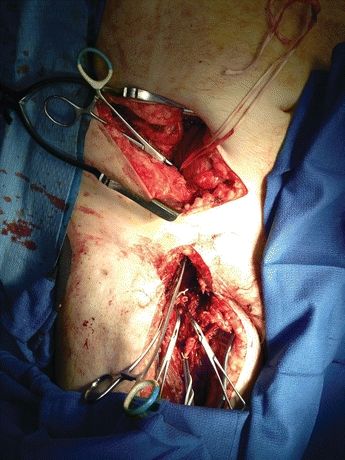
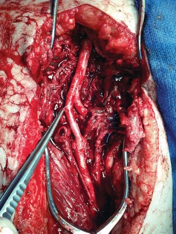
The patient was brought to the operating room, with pressure being applied to the groin. Both groins, abdomen, and legs were prepped, and patient was intubated. A “hockey stick” incision was made in the right lower abdomen 3 to 5 cm above the inguinal ligament. A standard retroperitoneal exposure of the external iliac artery was performed, and proximal control was achieved. The patient was heparinized, and distal control of the femoral artery was achieved distal to the area of rupture, obtaining control of the superficial and profunda femoral arteries (Fig. 1). Two sets of instruments were separately used for clean and contaminated portions of the procedure to minimize cross-contamination. Contralateral saphenous vein was harvested for the reconstruction. The infected common femoral, proximal superficial, and profunda femoral arteries were resected, and the area was widely debrided and curetted. Stat Gram stains were taken of the remaining margins and were debrided until they were negative for presence of bacteria. A distal external iliac artery to profunda femoris bypass was performed with reversed saphenous vein, and a jump graft was performed to the superficial femoral artery (SFA) (Fig. 2). Anastomoses were covered with a sartorius muscle flap. A vacuum-assisted dressing was applied. The patient was initially placed on broad-spectrum antibiotics, and these were adjusted to organism-specific antibiotics, which were given intravenously for 6 weeks.

FIGURE 1 Retroperitoneal exposure for proximal control (arrow). Standard femoral exposure for the distal control (double arrow). A: Clamp on eternal iliac artery. B: Profunda femoris artery. C: Superficial femoral artery.

FIGURE 2 External iliac to profunda and superficial femoral arteries bypass with the reversed greater saphenous vein.


