Intravascular Characterization of Vulnerable Coronary Plaque


Intravascular Characterization of Vulnerable Coronary Plaque


James A. Goldstein MD


Acute coronary syndromes (ACS) result from rupture of macrophage-rich, inflamed thin-capped fibroatheroma (TCFA) with superimposed thrombus formation (1, 2 and 3). Plaque disruption is thought to be a function of the interplay between factors that influence intrinsic plaque vulnerability and extrinsic forces that may precipitate rupture (1, 2, 3, 4, 5, 6 and 7). The invasive angiographic hallmark of ACS is a complex culprit plaque characterized by lesion irregularity, haziness, ulceration, contrast dye persistence within the plaque, intraluminal filling defect, and impaired flow (1, 4, 5). Although focal flow-limiting coronary stenoses are the targets for revascularization to relieve myocardial ischemia, angiographic observations support the concept that ruptured plaques most commonly arise from precursor “vulnerable plaques” (VPs) that, prior to disruption, are typically non-flow-limiting, asymptomatic, and would not be detectable by invasive flow interrogation with fractional flow reserve (FFR) or stress testing (2, 3, 6, 7 and 8). This VP hypothesis has led to a paradigm shift in thinking about the pathophysiology of coronary artery disease, with the focus no longer solely on the degree of arterial luminal narrowing (5, 6).

However, angiography is an insensitive tool that is only able to detect those plaques that have relatively gross plaque disruption and underestimates the magnitude of atherosclerotic burden, particularly in earlier-stage disease in which positive vascular remodeling may allow “normal” lumen caliber despite substantial vascular wall plaque (5, 6). Importantly, angiography has significant limitations in the precise delineation of plaque architecture and provides little or no information regarding plaque composition or its biological activity. This has led to a rapidly proliferating portfolio of invasive direct coronary imaging tools to better characterize atherosclerosis. This review will focus on those clinically available devices.


VULNERABLE PLAQUE: THE PRECURSOR LESION TO PLAQUE RUPTURE


Histopathologic Evidence

Autopsy studies of patients succumbing to acute myocardial infarction (MI) and to sudden coronary death suggest several underlying histological culprit plaque morphologies (1, 2, 8, 9), which thereby provide the foundation for the characterization of suspected VP thought to underlie ACS events. The most common morphology is an inflamed thin-cap fibroatheroma (TCFA), characterized histologically by: (a) thin fibrous cap (<65 µm), (b) large necrotic lipid pool, and (c) enzymatically active macrophages near or within the fibrous cap (1, 2 and 3, 8), (Figs. 9-1 and 9-2). These features are felt to predispose TCFA to rupture in response to internal and external biomechanical stresses (1, 2 and 3, 6). Following rupture, throm-bogenic plaque components exposed to luminal blood stimulate clot formation, which superimposed upon the plaque narrowing then progresse to an ACS event (1, 2, 3 and 4, 6, 7, 8 and 9). TCFAs are found most commonly within the most proximal 50 to 60 mm of the main coronary arteries (2, 9), and may be multifocal, especially in ACS cases (1, 4, 5, 9, 10, 11 and 12). Lesions with endothelial erosions and superficial calcium nodules may also be associated with thrombosis and unstable plaque events (13).


VULNERABLE PATIENT: IT’S NOT JUST THE PLAQUE, BUT ALSO THE MILIEU IT DWELLS WITHIN

The pathophysiologic factors underlying the transition from a stable lesion to a vulnerable plaque that subsequently undergoes disruption are not fully delineated. Likely mechanisms include local coronary and systemic inflammation, local coronary shear stress, alterations in lipid chemistry, status of blood thrombogenicity, and other factors (1). Regardless of the factors involved, disease transition may be reasonably considered analogous to the clinical “flares” characteristic of chronic inflammatory disorders (e.g., inflammatory bowel disease) in which a quiescent process smolders and then “flares.” The concept that such disease “flares” characterize ACS patients is supported by observations that demonstrate that, in comparison with those with chronic stable angina, unstable patients manifest evidence of multifocal plaque instability and multiple TCFAs, local and systemic inflammation, that persistent systemic inflammation predicts recurrent events and that in ACS cases are at higher risk for recurrent clinical instability arising from nonculprit lesions (3, 4, 5, 6, 7, 8, 9, 10, 11 and 12). In aggregate, these observations support the concept that plaque vulnerability and instability reflect pan-coronary destabilization processes associated with greater “disease activity.”


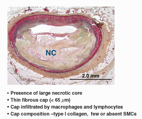
FIGURE 9-1 Accepted histological definition of TCFA/vulnerable plaque. (Courtesy James Muller, MD.)


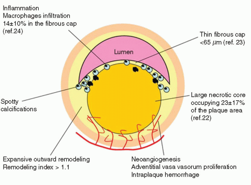
FIGURE 9-2 Typical morphologic traits associated with rupture prone plaques. (From: Vancraeynest D, et al. J Am Coll Cardiol. 2011;57:1961-1979, with permission.)

However, it must be emphasized that asymptomatic “stable” patients also are at risk for ACS events. Moreover, clinical symptomatic status does not necessarily reflect underlying plaque pathophysiology. The traditional view of “stable angina” is that this clinical entity typically reflects stable flow-limiting stenosis resulting from indolent, steady accretion of plaque. However, observations from pathologic and direct coronary imaging studies now document that some patients with clinically “stable” angina manifest evidence of plaque disruption (1, 2, 3, 4, 5, 6, 7, 8, 9, 10, 11 and 12). Such events could underlie the transition from an asymptomatic state to the onset of exertional angina, as plaque disruption facilitates rapid lesion progression, from a hemodynamically insignificant lesion to a flow-limiting stenosis. Thus, all patients with atherosclerosis are at risk for VP that may result in catastrophic complications, thereby offering a future opportunity to identify lesions at risk and potentially preempt disruption and its complications.


DEFINITIONS USED IN PLAQUE CHARACTERIZATION

First, it is essential to consider plaque characterization terminology and technology (14). The following definition of terms is offered: (a) “Unstable plaque,” the proximate cause of ACS characterized pathologically by a ruptured inflamed plaque with superimposed thrombus; (b) “Complex lesions,” the invasive angiographic hallmark of a frankly unstable plaque, characterized by haziness, fissuring, ulceration, filling defect, and impaired flow; (c) VP, a rupture-prone precursor of unstable plaque, linked pathologically to thin-capped fibroatheroma (TCFA); (d) “Culprit lesion,” the unstable plaque responsible for ACS; and (e) “Target” lesion, that plaque designated for revascularization. Unfortunately, although these definitions are widely accepted, these terms are often applied imprecisely or misused interchangeably.


PLAQUE CHARACTERIZATION TOOLS

An appreciation of the information necessary to precisely characterize plaques and fundamental data provided by plaque imaging technologies is essential. The ideal invasive tool for characterization of coronary plaque would provide a complete roadmap of atherosclerotic burden throughout the coronary tree and provide comprehensive per plaque analysis (Table 9-1, (6)).


Key Plaque Characterization Parameters

Key plaque characterization parameters include the following:


  • Architecture: Plaque volume, length, remodeling, eccentricity, impact on lumen area, fibrous cap thickness, evidence of disruption;


  • Physiology: Impact on coronary flow reserve;


    TABLE 9-1 Histologic Features of Suspected Vulnerable Plaquesa


    Type


    Features


    nflamed thin-cap fibroatheroma, most frequent type and present in 60% to 70% of ACS cases


    Necrotic core (lipid core) Thin cap <65µm nflammation


    Erosion site, present in 20% to 30% of events, more frequent in women and younger patients with ACS


    Increased vasa vasorum


    Expansive remodelling


    Increased plaque burden


    Intraplaque hemorrhage


    Calcified nodule, present in <3% of patients with ACS


    Spotty calcification


    Luminal narrowing


    Plaque with a mural thrombus not producing a significant stenosis


    Increased proteoglycans


    Extensive calcification protruding into lumen


    Considered to be a site of subsequent thrombosis because many plaque-causing events show repeated episodes of disruption and thrombosis


    ACS, acute coronary syndrome; Ml, myocardial infarction; PCI, percutaneous coronary intervention; TCFA, thin cap fibroatheroma


    aaFrom: Muller JE et al. J Am Coll Cardiol Imaging. 2010;3:881-891


  • Composition: Lipid-necrotic core, fibrous, hemorrhage, calcium, etc.;


  • Pathobiology: Presence of inflammation, neovascularization, fibrous cap metabolism, apoptosis.


INVASIVE TECHNIQUES FOR PLAQUE CHARACTERIZATION


Invasive Coronary Angiography: Limitations for Detection of Vulnerable Plaque

The invasive angiographic hallmark of ACS is a complex culprit plaque (Fig. 9-3), characterized by lesion irregularity, haziness, ulceration, contrast dye persistence within the plaque, intraluminal filling defect, and impaired flow (4, 5). These angiographic morphological features correlate with plaque rupture and thrombus by direct coronary imaging with intravascular ultrasound (IVUS) and at pathological examination (12, 15, 16 and 17). Observations from IVUS, and angioscopic and pathological studies document that the majority of ulcerated plaques are not sufficiently disrupted anatomically to be detected angiographically (2, 5, 8, 9, 10, 11, 12, 13, 14, 15

Only gold members can continue reading. Log In or Register to continue

Stay updated, free articles. Join our Telegram channel

May 28, 2016 | Posted by in CARDIOLOGY | Comments Off on Intravascular Characterization of Vulnerable Coronary Plaque

Full access? Get Clinical Tree

Get Clinical Tree app for offline access