Intracranial Occlusive Disease and Aneurysms: Influence on Carotid Revascularization Outcomes


Intracranial Occlusive Disease and Aneurysms


Influence on Carotid Revascularization Outcomes


Harry Ma, Adel M. Malek and William C. Mackey


Controversy has existed as to whether intracranial occlusive lesions or aneurysms alter the risk-to-benefit equation in patients undergoing carotid endarterectomy (CEA) or carotid stenting (CAS) by increasing the risk of perioperative stroke or by decreasing the long-term benefit. Two questions exist: (1) Should the presence of significant intracranial stenoses or aneurysms influence the decision to offer or withold CEA or CAS? and (2) With recent advancements in intracranial endovascular techniques, should we adopt a more-aggressive approach to incidentally discovered concomitant intracranial disease?


Intracranial Occlusive Disease


Intracranial occlusive lesions are found in approximately 20% to 40% of patients undergoing arteriography in preparation for CEA. The carotid siphon is the most common site of this disease. Mattos and colleagues identified siphon stenoses in 84 of 393 (21.4%) carotid arteries subjected to CEA, although stenoses exceeding 50% affected only 19 (4.8%) of these arteries. In the authors’ study, intracranial lesions were identified in 134 of 597 (22.4%) patients undergoing CEA. Carotid siphon occlusive disease was present in 90 patients (15.1%), but high-grade (more than 80%) siphon lesions were present in only 11 cases (1.8%). Schuler and coworkers identified siphon lesions of at least 20% in 44 of 91 (48%) carotid arteriograms, and in 14 of 91 (15.4%) the stenosis exceeded 50%. Keagy and associates and Roederer and colleagues identified more than 50% siphon stenosis in 17% and 9% of arteriographically visualized carotids, respectively. Rouleau’s group retrospectively reviewed 672 patients who underwent cerebral angiography and found 65 patients (9.7%) with siphon stenoses of 50% or greater, but only 37 patients (5.5%) had an ipsilateral internal carotid artery stenosis of 70% or greater. Twenty-six of the 37 patients with tandem lesions underwent CEA, resulting in only two minor postoperative complications.


It may be concluded that carotid siphon disease and other intracranial occlusive lesions are often found in patients undergoing evaluation for CEA. High-grade stenoses of the siphon are unusual. In current practice the true incidence and significance of intracranial disease in conjunction with extracranial carotid stenosis is likely to be underreported given that many CEAs are performed based solely on data obtained from duplex ultrasonography rather than complete cerebral angiograms.


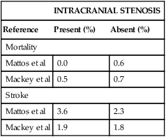
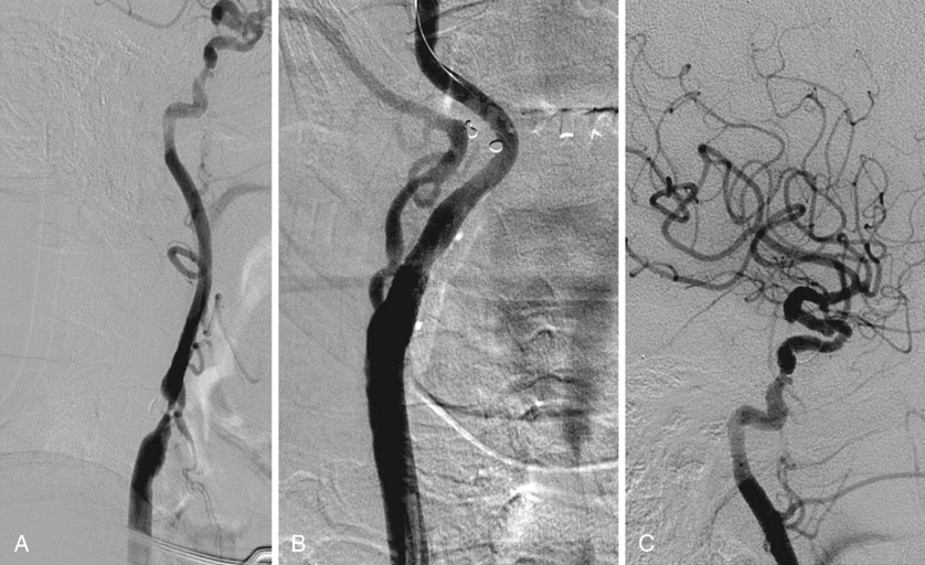
Perioperative (30 day) stroke and death rates are not statistically significantly increased in CEA patients with intracranial occlusive lesions (Table 1). In a subgroup of patients with more than 50% siphon stenosis, Mattos and coworkers found a perioperative stroke rate of 1 in 19 (5.3%), which was not statistically significantly different from the stroke rate noted in patients without siphon disease or with mild (20%–49%) siphon lesions. This is similar to the findings of Schuler and associates, who noted perioperative stroke rate of 7.1% in patients with more than 50% ipsilateral siphon stenosis. In addition, they found a stroke rate of 2 in 18 (11.1%) in patients undergoing endarterectomy in whom the siphon stenosis exceeded the bifurcation stenosis. Although this was greater than the perioperative stroke risk (4.1%) noted in patients with normal or less severely diseased siphons, the difference was not statistically significant.


Data on CAS in patients with tandem lesions consist of case reports and small series. Tsutsumi and colleagues reported successful treatment of two patients with simultaneous stenting of both intracranial and extracranial carotid stenoses. Both of these patients had greater than 70% stenosis of their ICA and greater than 80% stenosis of the ipsilateral intracranial carotid artery. Cohen and colleagues reported a series of eight patients with symptomatic tandem lesions treated by sequential stent-assisted angioplasty. There were no strokes, transient ischemic attacks (TIAs), or myocardial infarctions (MIs) noted in the perioperative period, and there were no neurologic events and no in-stent restenosis noted at 6 months. In a larger multicenter review, Siddiqui and coauthors assessed the rate of stroke or death in patients undergoing CAS in patients with and without tandem lesions. Similar to the results for CEA, patients with tandem lesions had a trend toward increased perioperative and midterm stroke and/or death rates, but this trend did not reach statistical significance.


Perioperative stroke risk, then, may be slightly increased in patients with intracranial occlusive lesions, especially in those unusual patients with more than 50% siphon lesions. However, the added risk is slight and in these small series did not achieve statistical significance. Of course, because of the small numbers of subjects in these series a type II statistical error is likely. Unfortunately, no larger series or meta-analyses are available. Even less is known about the influence of intracranial stenoses on CAS results, though initial studies suggest a similar trend.


The long-term stroke prevention afforded patients by CEA is only slightly diminished by the presence of intracranial occlusive lesions. In neither the Mattos and associates nor the Mackey and associates studies did the differences in late Kaplan Meier–derived stroke-free rates approach statistical significance. Similarly, Roederer and colleagues found no correlation between the likelihood of symptoms recurring and the presence or severity of siphon disease in carotid endarterectomy patients.


The influence of intracranial occlusive disease on long-term survival after CEA remains unclear. Mattos and colleagues found a statistically significant stroke-free survival disadvantage (49.1% vs. 75.3% at 7 years; p = .04) in patients with siphon lesions. In the Mackey study, life table stroke-free survival was not different in patients with and without siphon stenosis (p = .75). Review of the demographic and risk-factor data from these two studies reveals no explanation for these discrepant results. Most of the excess mortality in Mattos’s patients with siphon disease occurred more than 5 years after CEA, and that study had relatively small numbers of patients followed beyond 5 years. These factors raise the possibility that the apparent increase in late mortality in Mattos’s siphon stenosis patients is artifactual. In all studies the most common cause of death in patients with and without intracranial lesions was myocardial infarction, not stroke.


Intracranial occlusive lesions, then, appear to have only minor influence on perioperative mortality and stroke morbidity and negligible influence on long-term stroke-free rates in CEA patients. Most intracranial lesions found in CEA candidates are in the carotid siphon and are less than 50% stenoses. Their lack of influence on late stroke morbidity suggests that these lesions remain stable. The benign natural history of carotid siphon lesions may be related to their morphologic characteristics. Unlike plaques at the carotid bifurcation, which calcify as they degenerate, siphon plaques can calcify early in their progression. This early calcification involves only the media and elastic lamina, is not accompanied by other morphologic characteristics of progressive atherosclerosis, and does not appear to correlate with the presence or severity of atherosclerosis elsewhere. Fisher and coworkers postulated that diffuse calcification of the media and elastic lamina of the carotid siphon might actually retard local progression of more typical obstructive atherosclerosis. Many if not most of the nonstenosing plaques noted in studies of intracranial occlusive lesions may be related to diffuse medial calcification and therefore be more stable than the typical atherosclerotic plaque.


Because intracranial occlusive lesions have only minor effects on perioperative mortality or stroke risk or on long-term stroke-free rates, most patients with appropriate carotid bifurcation pathology and intracranial occlusive lesions are acceptable candidates for CEA or CAS. Thus a symptomatic patient with a compelling bifurcation lesion and a smooth intracranial stenosis (Figure 1A) may subsequently undergo CAS or CEA (Figure 1B and C). The presence of mild to moderate intracranial occlusive disease in this setting should not influence a decision to intervene.


Stay updated, free articles. Join our Telegram channel

Aug 25, 2016 | Posted by in CARDIOLOGY | Comments Off on Intracranial Occlusive Disease and Aneurysms: Influence on Carotid Revascularization Outcomes

Full access? Get Clinical Tree

Get Clinical Tree app for offline access