LQT2 is the second most common loci for LQTS (35% to 40%) and stems from mutations in the KCNH2 gene encoding the alpha (HERG) subunit of the potassium channel responsible for the IKr current. The T waves in LQT2 are usually low amplitude and bifid (see Fig. 8.1B).
LQT3 arises from gain of function mutations in the SCN5A that encodes for the rapidly inactivating sodium channels NaV1.5. The ECG in LQT3 shows long isoelectric ST segments with a late-appearing T wave (see Fig. 8.1C).
ELECTROCARDIOGRAM AS USED IN DIAGNOSIS
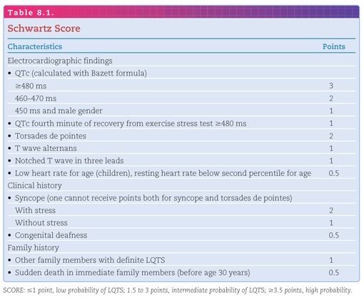
It is important to remember that a prolonged QT interval alone is not sufficient to make the diagnosis of LQTS; an increased risk of SCD is necessary. To assist with the diagnosis of LQTS, a diagnostic score has been created, known as the International Long QT Score or “Schwartz score” (Table 8.1). Although a score ≥3.5 makes the diagnosis of LQTS more likely, it also is not diagnostic.8
As with LQTS, short QT syndrome (SQTS) is a disorder of repolarization, in this case associated with more rapid repolarization and therefore a short QT interval. Again, like LQTS, this condition can be congenital or acquired. Acquired causes of short QT include hyperthermia, hyperkalemia, hypercalcemia, acidosis, and changes in autonomic tone. Congenital SQTS is much more rare than LQTS, with <100 cases reported worldwide. SQTS is defined by the presence of an abnormal QT interval (<300 milliseconds) and an increased risk of ventricular arrhythmias and SCD. Not surprisingly, the genes currently associated with SQTS are involved in the repolarization phase of the cardiac action potential. These functional mutations lead to a gain-of-function in the three voltage-gated potassium channel genes: KCNH2, KCNQ1, and KCNJ2. This gain of function results in an increased efflux of potassium from the cell during the repolarization phase and a shortening of the action potential.9
ELECTROCARDIOGRAPHIC CHARACTERISTICS
QT Interval
Similar to LQTS, the QT interval in a population follows a normal distribution, thus there will be many “normal” patients in the general population with a short QT interval <360 milliseconds. However, patients with very short QT intervals (QTc <330 milliseconds in males and QTc <340 milliseconds in females) should be considered for SQTS even if they are asymptomatic because QT intervals this short are quite rare.
T-Wave Morphology
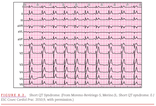
Most SQTS patients have an absent ST segment, with the T wave beginning immediately after the S wave. The T wave also often appears peaked and narrow (Fig. 8.2).
ELECTROCARDIOGRAM AS USED IN DIAGNOSIS
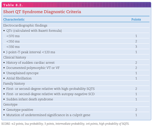
Similar to LQTS, a prolonged QT interval alone is not sufficient to make the diagnosis of SQTS; an increased risk of SCD is necessary. To assist with the diagnosis of SQTS, a diagnostic score has been created (Table 8.2).9 Although a score ≥4 makes the diagnosis of SQTS more likely, it is not sufficient to make the diagnosis.
The Brugada pattern was first reported in 1953, but it was not until ECG pattern was associated with SCD in 1992 that it became a recognized clinical syndrome.
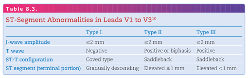
The Brugada pattern is the hallmark of the syndrome and has a characteristic pattern on ECG consisting of a pseudo–right-bundle-branch block (RBBB) and persistent ST-segment elevation in leads V1 to V3.10
Since the original description by Brugada et al,11 there have been three main patterns described: (Table 8.3, Fig. 8.3)

Stay updated, free articles. Join our Telegram channel

Full access? Get Clinical Tree


