Infrainguinal Endovascular Reconstruction: Technique and Results

Chapter 28 Infrainguinal Endovascular Reconstruction


Technique and Results


Management of infrainguinal arterial occlusive disease continues to move away from open surgery and toward percutaneous procedures, and the number of percutaneous options is growing rapidly. This chapter focuses on standard techniques for arterial access, diagnostic imaging, lesion crossing, and options for treating occlusions and stenoses of the femoropopliteal and tibial circulation.


Patient Selection and Preoperative Imaging


Infrainguinal occlusive disease can usually be diagnosed by history and physical examination. Confirmatory studies are usually performed—either duplex mapping or magnetic resonance angiography—in order to plan the therapeutic approach and limit the amount of contrast required for arteriography. Diagnostic arteriography does not currently exist in many practices, because most patients do not undergo arterial access unless there is an intention to treat. Occasionally, what initially appeared to be a lesion appropriate for angioplasty based on duplex scanning or magnetic resonance angiography turns out to be more complex, and only an arteriogram is obtained.


Infrainguinal occlusive disease can be classified by its morphology, which assists in determining which patients are best managed with endovascular intervention and which ones require surgery. The TransAtlantic Inter-Society Consensus (TASC) classification (Box 28-1) and others have defined disease morphology in an effort to clarify the issue of lesion severity.1 The general concept is that endovascular techniques are preferred in patients with less severe forms of disease and among those with shorter life expectancies or greater periprocedural risk factors. Conversely, open surgical approaches have a better risk-benefit profile in patients with fewer medical comorbidities or more severe forms of disease, such as long-segment occlusions, in which endovascular procedures are less durable. The recommendation from the TASC group is that type A lesions be treated with endovascular intervention, type D lesions be treated with surgery, and types B and C lesions be treated with either, depending on the patient’s comorbidities, general medical condition, expected longevity, and availability of conduit for bypass.


Over the past decade, many practices have seen a steady movement toward the use of endovascular techniques for infrainguinal arterial occlusive disease, to the point that some approach all patients with an “endovascular first” strategy with open surgical bypass reserved for failures of endovascular therapy. However, some lesions or disease patterns continue to be best treated with open surgery, such as common femoral disease or long-segment femoropopliteal occlusions with distal tibial reconstitution, although even these difficult disease patterns can be treated with some of the newer percutaneous modalities, such as laser or excisional atherectomy.


Approaches


Percutaneous intervention for infrainguinal occlusive disease is usually performed through the contralateral femoral artery using an up-and-over approach or through the ipsilateral femoral artery using an antegrade approach (Table 28-1). Infrainguinal interventions can also be performed through the brachial artery, but this approach is rarely required and may be more challenging because of the longer distances involved and the higher likelihood of access-related complications. The primary advantages of the up-and-over approach, which is most commonly used, are the following: an aortogram with runoff can be easily converted to endovascular therapy; it permits evaluation of the inflow aortoiliac arteries before treatment of infrainguinal lesions; only a simple retrograde femoral puncture is required; and it facilitates selective catheterization of the superficial femoral artery (SFA) orifice and treatment of proximal SFA lesions, which can be difficult via the ipsilateral antegrade approach. Furthermore, puncture site management is contralateral to the intervention site rather than proximal to it. The antegrade approach may be used for better guidewire and catheter control in infrapopliteal intervention and also in patients who have contraindications to the up-and-over approach. The likely approach is determined before the procedure in order to facilitate room setup and the availability of supplies, but both groins are always prepared in case an alternative approach is required during the procedure.


TABLE 28-1 Comparison of Approaches to Infrainguinal Interventions: Up-and-Over versus Antegrade


  Up-and-Over Approach Antegrade Approach
Puncture Simple retrograde femoral More challenging, less working room
Catheterization Challenging with tortuous arteries, narrow or diseased aortic bifurcation; easier to catheterize SFA when going up and over femoral Entering SFA from antegrade approach requires proximal puncture and selective catheter
Guidewire and catheter control Fair Excellent
Catheter inventory More supplies needed Minimal, shorter catheters
Specialty items Up-and-over sheath, long balloon catheters None
Indications Proximal SFA disease, CFA disease ipsilateral to infrainguinal lesion, obesity Infrapopliteal disease, patients with contraindication to up-and-over approach

CFA, Common femoral artery; SFA, superior femoral artery.


From Schneider PA: The infrainguinal arteries—advice about balloon angioplasty and stent placement. In Endovascular skills, ed 2, New York, 2003, Marcel Dekker, p 316.


Platforms


Percutaneous intervention in the infrainguinal circulation can be achieved with either 0.035-inch or 0.014-inch platforms, using sheath sizes ranging from 4 to 7 French. Most diagnostic angiograms and interventions are begun with a standard 0.035-inch platform. This platform has several advantages: the guidewires and catheters are easy to handle, the inventory is usually readily available, the fluoroscopic visualization of these larger-caliber devices is simpler, and the larger guidewires and catheters are often more helpful than the smaller diameter devices when crossing chronic or heavily calcified occlusions. However, there are also some significant disadvantages of 0.035-inch systems: the larger-caliber guidewires and catheters may not easily cross critically diseased segments and may be more prone to cause arterial damage; at longer distances, these catheters lose their “pushability” because of high friction; and in small arteries such as tibial vessels, the standard platform devices may be too big. In addition to their smaller crossing profiles, the smaller 0.018- and 0.014-inch platforms tend to be more trackable in small, tortuous vessels at distant locations from the access site, especially when using monorail (rapid-exchange) systems with longer guiding sheaths or catheters. Most of the coronary devices are on a 0.014-inch or 0.018-inch platform, as are current atherectomy systems; therefore the array of available balloon catheters and stents is much broader with this system. Monorail balloon catheters permit better pushability because the friction of the guidewire on the balloon catheter lumen occurs over a much shorter distance than with coaxial balloon catheters. Monorail systems have the additional advantages of greater ease of use (especially with a single operator), shorter required guidewire lengths, and less guidewire movement during catheter exchanges.


Technique


Up-and-Over Approach


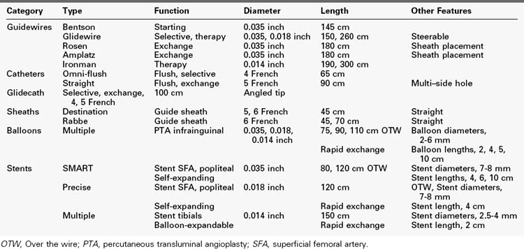
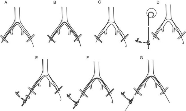
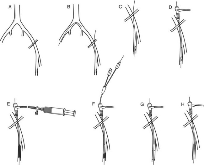
Supplies required for an up-and-over approach are listed in Table 28-2. This approach requires longer guidewires, catheters, and sheaths than the antegrade approach. A standard retrograde common femoral artery puncture is performed contralateral to the symptomatic side. A floppy-tipped guidewire is passed into the aorta. A hook-shaped, multi–side-hole flush catheter, such as a 65-cm Omni-flush (AngioDynamics), is passed into the aorta, and an aortoiliac arteriogram is obtained. If bilateral runoff is required, it can be performed at that time with the catheter head placed in the infrarenal aorta. When only unilateral runoff on the symptomatic side is indicated, the catheter is passed over the aortic bifurcation, and lower extremity arteriography is performed. After evaluating the infrainguinal lesions, determining that the aortic bifurcation can accommodate an access sheath, and deciding that the up-and-over approach is best, an up-and-over sheath is placed (Figure 28-1).


The aortic flush catheter is withdrawn to the aortic bifurcation, and its tip is rotated toward the contralateral side to direct the guidewire into the contralateral iliac artery. The advancing guidewire, usually a steerable, angled-tip Glidewire (Terumo Medical, Tokyo, Japan), must be steered into the external iliac artery and then into the infrainguinal arteries. From this approach, the guidewire usually tends to select the contralateral internal iliac artery if there is tortuosity of the iliac system. The guidewire also tends to select the SFA rather than the deep femoral artery. Either of these destinations for the guidewire is satisfactory from the standpoint of sheath placement, as long as the guidewire is well anchored distal to the groin. The catheter is advanced over the bifurcation, and a stiffer exchange-length (260 cm) guidewire (Stiff Angled Glidewire [Terumo Medical], Supra-Core Wire [Abbott Vascular, Abbott Park, Ill.] or Rosen Wire [Cook Medical]) is placed. The tip of the exchange guidewire should be distal to the groin as far as it will easily travel. If there is a proximal SFA lesion that is planned for treatment, the guidewire is usually directed into the deep profunda femoris artery. If there is excessive iliac tortuosity, an Amplatz Superstiff guidewire (Cook Medical) may be required.


Dilators are used to enlarge the arteriotomy. Because dilators are sized by their outside diameter and sheaths are sized by their inside diameter, if a 6-French sheath is planned for placement, the tract should be dilated using a 7-French dilator. The guide sheath is placed on the guidewire; when a curved sheath is used, it is oriented such that its curved end is pointing toward the contralateral side and the side arm of the sheath is on the side of the operator. The sheath is advanced over the guidewire using fluoroscopy. Passage over a narrow or diseased aortic bifurcation is performed with care and patience. The sheath is advanced to its hub, if possible. It is important to remember that the tip of the dilator extends beyond the radiopaque marker on the end of the sheath tip for a short distance, and injury to the arteries can occur if this dilator advances beyond the end of the guidewire. The tip of the sheath will generally terminate somewhere between the mid external iliac artery and the very proximal SFA, depending on the height of the patient. Heparin is usually administered (100 U/kg) as the sheath is placed, and an activated clotting time (ACT) is checked (goal ACT > 250 seconds).


Figure 28-2 demonstrates the steps required for infrainguinal intervention using an up-and-over approach. The exchange guidewire is replaced with a steerable 260-cm Glidewire (Terumo Medical). The diseased infrainguinal segment is road-mapped through the side arm of the sheath, and the Glidewire is used to cross the lesion to be treated. Lesion crossing is facilitated by supporting the Glidewire with a hydrophilic catheter, such as a 5-French Angled Glidecath (Cook Medical) or Quick Cross catheter (AngioDynamics). If treatment of tibial lesions is planned, a 5-French, 100-cm-long catheter is advanced into the distal popliteal artery, and road mapping is performed through this catheter. A low-profile guidewire, usually an 0.014-inch hydrophilic wire such as an Asahi Grandslam (Abbott Vascular) is used to cross the tibial lesions, and the 5-French diagnostic catheter is exchanged for a 135-cm–long, 0.014-inch Quick Cross catheter (AngioDynamics) to support the wire. Interval arteriography may be performed through the side arm of the sheath or through the selective catheter using a Tuohy-Borst adapter during attempts at lesion crossing. Once the lesion has been crossed, the supporting catheter is advanced down to the level of the reconstituted artery, the wire is removed to assess for blood return from the catheter lumen, and confirmatory angiography is performed to demonstrate the intraluminal position of the catheter.


Antegrade Approach


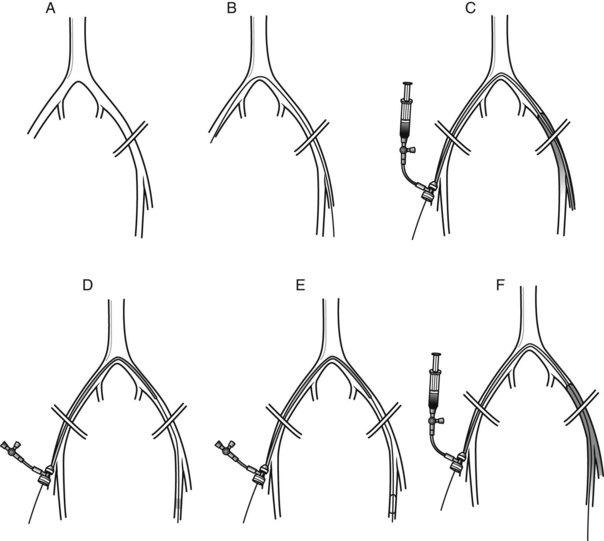
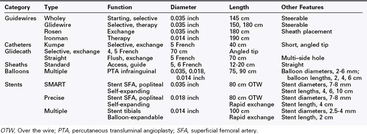
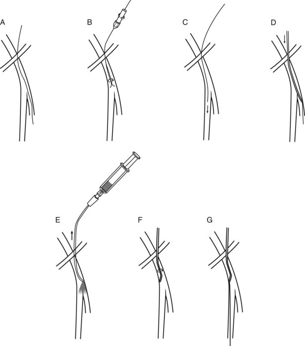
Supplies required for an antegrade approach are listed in Table 28-3. An antegrade common femoral artery puncture is performed ipsilateral to the symptomatic side. This approach is well suited to patients who have normal aortoiliac inflow, especially if tibial angioplasty is required or if there is a need to limit the use of contrast material. The puncture should be performed as proximally as possible along the common femoral artery to leave some working room between the puncture and the origin of the SFA. A steerable guidewire, such as the Wholey guidewire (Mallinckrodt, Hazelwood, Md.), is used; the shaft of this guidewire is more supportive for catheter and sheath passage than a Glidewire. The Wholey guidewire can often be steered anteromedially into the SFA. If not, the guidewire is advanced into the deep femoral artery, and an angled-tip catheter is placed over it (Figure 28-3). The image intensifier is placed in the ipsilateral anterior oblique position to open the femoral bifurcation, and the catheter is withdrawn enough to perform road mapping by refluxing contrast material into the SFA. The catheter is used to steer the guidewire into the SFA.


If the lesion is in the proximal to mid SFA, the artery is road mapped using the catheter, and the guidewire is advanced across the lesion (Figure 28-4). The same guidewire can be used for sheath placement. If the lesion is more distal in the artery, the guidewire is advanced without crossing the lesion, and the sheath is placed. The sheath size required may be 4, 5, or 6 French, depending on the platform used and treatment modality desired. An appropriately sized dilator is used to enlarge the arteriotomy before sheath placement.


After the sheath is placed, femoral arteriography is performed through its side arm. Heparin is administered (100 mg/kg) and the ACT is assessed. Standard-length, 180-cm, 0.035-inch guidewires may be used for lesions above the knee. Longer 260-cm, 0.014- or 0.018-inch guidewires are used for infrageniculate intervention, especially in tall patients. As with the contralateral approach, the lesion is then evaluated angiographically, and a steerable guidewire and supportive hydrophilic catheter is used to cross the lesion. Arteriography is repeated after the guidewire is across the lesion to be certain that the guidewire is in the distal artery and not in a perigenicular collateral. The lesion is then treated with the operator’s preferred treatment modality, and the completion arteriography is performed through the sheath while maintaining guidewire control until the results are assessed.


Treatment Modalities


Approach, sheath placement, and lesion crossing are performed similarly regardless of the ultimate treatment modality that is chosen. Once the lesion has been crossed and confirmation of true-luminal reentry has been ascertained, the operator can choose from an increasing number of percutaneous treatment modalities. Although much of this decision is based on operator preference and comfort level, certain modalities are better suited for specific disease patterns.


Balloon Angioplasty


Balloon angioplasty has been used since the 1980s and has the advantage of being technically simple and quick to perform, thus minimizing contrast usage and radiation exposure. Most superficial femoral and popliteal artery lesions are treated with balloons ranging from 4 to 6 mm in diameter, and a number of vendors provide balloons in lengths up to 200 mm. Tibial arteries range from 1 to 4 mm in diameter, but most are treated with balloons ranging from 2 to 3.5 mm in diameter and up to 150 mm in length. Increased balloon lengths allow long-segment lesions to be treated faster by minimizing the number of inflations and reducing the number of times a recently treated area is subjected to stagnation of blood flow because of a balloon inflation proximal or distal to it. Most modern angioplasty balloon catheters are relatively noncompliant in nature, thus ensuring that the balloon diameter varies little, even at high-pressure inflations.


Catheter length must be anticipated before selecting the balloon. Most of the standard 0.035-, 0.014-, or 0.018-inch platform balloons are on shafts that are either 75 to 80 cm long or 120 to 150 cm long. A 75-cm balloon catheter shaft passed up and over the bifurcation will reach anywhere from the common femoral artery to the distal SFA, depending on the patient’s height. An estimation of the distance to the lesion can be obtained by using a 75-cm–long straight exchange catheter for guidewire exchanges. The up-and-over sheath also provides clues because its length is known.


When performing angioplasty of an infrainguinal lesion, the balloon catheter is passed over the guidewire and into the lesion. The location of the lesion may be marked using road mapping or an external marker. The balloon is then inflated until the waist on the balloon profile is resolved, which generally requires inflations up to 6 to 10 atm. The duration of balloon inflation is anywhere from a few seconds to several minutes. If the waist caused by a lesion fails to resolve even at high inflation pressures, this can sometimes be remedied by exchanging a long balloon for a short (2 cm long) balloon which is then centered directly at the waist and inflated to the balloon’s rated burst pressure. Following treatment, the completion arteriogram is obtained through the side arm of the sheath. Balloon angioplasty of the superficial femoral and popliteal arteries almost always produces some evidence of dissection on completion arteriography. In this setting, deciding which patients require a stent may be challenging, though stents are generally used in patients with flow-limiting dissections, large spiral dissections, or residual stenoses or dissections resulting in greater than 30% reduction in flow lumen diameter.

< div class='tao-gold-member'>

Stay updated, free articles. Join our Telegram channel

Jul 1, 2016 | Posted by in CARDIOLOGY | Comments Off on Infrainguinal Endovascular Reconstruction: Technique and Results

Full access? Get Clinical Tree
Get Clinical Tree app for offline access