Intravenous drug users generally avoid regular physician contact, and care is often not sought until the infectious process is in its advanced stages. In our patients, the average delay between onset of symptoms and vascular surgical consultation was 17 days. The analgesic effect of continued opiate use contributes to prehospital delays in management. Self-administration of antibiotics by drug abusers is common and can temporarily mask or control the infectious symptoms. It is not unusual for the patient to persist with injections into the site.
A tender groin mass, almost invariably present, is pulsatile in 50% of patients. When sought, a bruit is evident in more than 90%. An open punctate skin lesion draining serosanguineous fluid is characteristic. In substance abusers, the contralateral groin might also exhibit a depressed circular scar, indicating bilateral self-injection. Other stigmata of chronic drug use may be present; however, an occasional patient comes to the hospital with evidence of groin injection only. Evidence of distal embolization is present in 5% to 10% of patients. Because this aneurysm is not readily distinguished from abscesses, an ill-advised incision and drainage is a trap for the unwary. Rupture occurs in 5% to 15%. The infected aneurysm is often not recognized in the early stages of the process, and extensive damage may be present at the first contact with vascular surgical personnel.
Systemic infection and bacteremia are common. Staphylococcus aureus is recovered most often. Pseudomonas aeruginosa is common, and unusual microbiologic organisms are also cultured. Reddy and coworkers observed 35 of 54 patients (65%) to have pure Staphylococcus aureus infections, and 17 of those had methicillin-resistant S. aureus (MRSA). Other communicable infectious diseases are also endemic in the intravenous drug user population.
Adjacent structures are commonly involved by the pseudoaneurysm that has progressed for several weeks. Femoral nerve compression resulted in substantial morbidity with little subsequent recovery in our patient population. This alone accounted for major disability following treatment of the infected aneurysm. Likewise, with chronic injection, localized thrombosis of the femoral vein is common. Inability to extend the hip fully indicates extension of the infection deep into the thigh, and such patients comes to the hospital with their hip flexed and externally rotated.
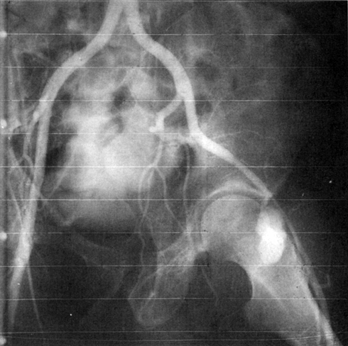
Arteriography distinguishes abscess from pseudoaneurysm and localizes the arterial defect anatomically (Figures 1 and 2). The anatomic location of the aneurysm determines which and how many arteries must be ligated, and it may be useful in predicting the probability of limb ischemia. Occasionally an aneurysm originates from a branch of the profunda femoris artery (see Figure 2). Because occult thromboembolism may be an important determinant of the final outcome of the limb, visualization of the distal arterial runoff becomes important if bypass is considered. Although arteriographic information can substantially alter operative management, information derived from computed tomography angiography (CTA) is often adequate and can obviate the need for this additional examination.
Only gold members can continue reading.
Log In or
Register to continue
Related
![]()


