Imaging for Structural Heart Disease


Imaging for Structural Heart Disease


Mazen Abu-Fadel MD, FACC, FSCAI


Recent technological advances and a cascade of procedural techniques and devices have produced a surge in the number and type of procedures performed for structural heart disease (SHD).

SHD interventions range from well-established procedures such as atrial septal defect (ASD) closure and balloon valvuloplasty to investigational techniques such as mitral valve clipping and left atrial appendage (LAA) occlusion. The number of catheter-based corrective procedures is growing rapidly mainly because of the aging population and the outcomes of recent and ongoing clinical trials investigating patients with SHD (1). Parallel to the development of these procedures and techniques, the role of imaging in the cardiac catheterization laboratory has become an integral and vital part of such interventions. Different imaging modalities, especially echocardiography, are crucial to help plan, guide, and optimize procedural success and to detect and treat complications as needed.

The first part of this chapter reviews the rationale and use of different imaging modalities as an integral part of SHD interventions. The second part discusses a general overview of the use of imaging in specific cardiac structural interventions.


IMAGING MODALITIES FOR SHD


Fluoroscopy

Despite the growth and development of multiple noninvasive imaging techniques, fluoroscopy remains an integral part of any catheterization laboratory interventional procedure. Although simple procedures such as patent foramen ovale (PFO) and small ASD closure have been done under echocardiographic guidance only (especially in pregnant women), fluoroscopy is needed for catheterbased interventions. Both fluoroscopy and angiography have an inherit limitation in the accurate diagnosis of SHD. X-ray equipment provides a two-dimensional (2-D) representation of a threedimensional ??????(3-D) structure. Even with dual plane catheterization laboratories, special information may still be lacking, and cardiac soft tissues cannot be identified. Moreover, ionizing radiation use increases the risk of injury to the patient and the operator, especially in long complex cases.

Owing to the limitations of fluoroscopy and the development of other reliable and less risky imaging modalities with superior soft tissue images and better special resolution, SHD interventions are currently being performed with much less need for live fluoroscopy.


Echocardiography

Echocardiography is essential as it plays a crucial role in identifying patients suitable for the procedure, provides intraprocedural guidance, and remains the primary modality for postprocedure followup. The demands on the echocardiographer to interpret and guide catheter-based interventions for SHD are much more complex than interpreting a routine echocardiogram. Consequently, interventional cardiologists—if not well trained—will require the assistance of a skilled echocardiographer as part of the interventional SHD team.

The major advantage of echocardiography over other imaging modalities such as computed tomography (CT) and magnetic resonance imaging (MRI) is its mobility and ease of use. In addition, it does not require any contrast agent, and does not emit ionizing radiations. It can produce real-time 2-D and 3-D images, and has the ability to detect and diagnose complications in real time during the procedure, which helps for early correction and treatment. Transthoracic echocardiography (TTE), transesophageal echocardiography (TEE), and intracardiac echocardiography (ICE) are the three modalities widely used for SHD interventions. The advantages and limitations of each are listed in Table 38-1.


Transthoracic Echocardiography

TTE is noninvasive and widely available. It does not require patient sedation or general anesthesia. Newer systems are small and portable and provide adequate 2-D imaging and Doppler capabilities to assist in a number of procedures, including mitral balloon valvuloplasty, alcohol septal ablation, atrial septostomy, and others (2). TTE images may be challenging to acquire with the patient supine on the catheterization table, and usually the probe and the operators’ arm may be in the fluoroscopic field, and thus simultaneous images may not be obtainable (3).


Transesophageal Echocardiography

Currently TEE is the standard imaging technique in many centers that perform catheter-based interventions (3). TEE offers superior imaging quality when compared with TTE. It provides direct assessment of intraprocedural anatomy and physiology, shows the relationship between catheters/devices and cardiac structures, and helps guide interventions and diagnose complications. TEE imaging does not interfere with the operative field and rarely interferes with the fluoroscopic field. One of the most important limitations of TEE includes the need for deep sedation or general anesthesia with endotracheal intubation. In addition, long cases may predispose the TEE probe to overheat, causing burns in the esophagus (2).

On the other hand, TEE has the capability to provide real-time 3-D imaging. This capability has overcome the limitations of other 3-D imaging modalities such as CT and MRI that have to acquire images prior to the procedure and process them offline to obtain 3-D reconstructions. Real-time 3-D TEE (RT 3D TEE) can provide immediate and online images of cardiac structures, including mobile structures; superior spatial relationship; and alignment of catheters and devices to the cardiac defects. These defects often have a complex morphology that is suboptimally visualized by 2-D imaging, including TEE (4). In addition, 3-D TEE provides en face views of cardiac structures, valves, and surrounding tissues,
which has proved to be of great value in some procedures such as repair of paravalvular leaks (5). RT 3D TEE has a steep learning curve and requires effective communication between the echocardiographer and the interventionalist (1).


TABLE 38-1 Advantages and Limitations of Echocardiographic Imaging Techniques


Advantages


Limitations


Use for Guidance


TTE


Easy and rapid access ++


Limited acoustic windows ++


TAVI +


Good image quality (harmonic imaging)


Interaction with the sterile field


Doppler capabilities


Not usable simultaneously with fluoroscopy


RT 3D capabilities


TOE


High-resolution imaging +++


Semi-invasive


Transseptal puncture +++


Doppler capabilities


Discomfort and aspiration risk in conscious


BMV+++


Multiplane imaging


patients


TAVI ++


Accurate assessment of posteriorly situated cardiac structures (interarterial septum,
LAA, mitral and aortic valve)


Usually requires general anaesthesia and endotracheal intubation ++


Other complex percutaneous


procedures +++


Absence of interference with the operative field


RT 3D capabilities +++


ICE


High-resolution imaging +++


Far field imaging


Trans septal puncture +++


Doppler capabilities and four-way steerability (phased array ICE)


Rare vascular complications (hematomas, venous thrombosis)


BMV++ Percutaneous ASD and PFO closure +++


Accurate assessment of interatrial septum, left atrium, pulmonary veins


Arrhythmias Learning curve


TAVI +


No need for general anaesthesia


Additional cost ++


Reduced length of procedure and fluoroscopy time


3D (not real time)


ASD, atrial septal defect; BMV, balloon mitral valvuloplasty; ICE, intra cardiac echocardiography; LAA, left atrial appendage; PFO, patent foramen ovale; RT 3D, real-time three dimensional; TAVI, transcatheter aortic valve implantation; TTE, Transthoracic echocardiography.


From: Brochet E, et al. Heart. 2010;96(17):1409-1417, with permission


Intracardiac Echocardiography

ICE has demonstrated an excellent potential to guide and monitor catheter-based interventions. The image quality of ICE is comparable or at times superior to TEE. This technology is widely used for a broad variety of SHD and electrophysiological procedures. Even though ICE is an invasive procedure, one of its major advantages is the elimination of the need for general anesthesia and endotracheal intubation as in TEE (2).

The most commonly used ICE catheters come in 8- and 10-F shafts with a transducer frequency that ranges from about 5 to 10 MHz and has Doppler and color flow capabilities. The catheter is attached to a control handle that helps steer it in four different directions (anterior-posterior and left-right) using two different knobs. The third knob locks the position of the catheter in place once the desired image is displayed (6). The probe is advanced through a separate sheath in the ipsilateral or contralateral femoral vein into the right atrium. Generally, a long sheath from the femoral vein is advanced into the inferior vena cava, and then the ICE catheter is advanced under fluoroscopic guidance because of its rigidity and its blunt tip that may get wedged in venous branches, causing potential injury to vital organs and bleeding (7). When the catheter reaches the right atrium, the standard starting view, called the “home view,” shows the right atrium, right ventricle, tricuspid valve, anterior part of the anterior septum, and the right ventricular inflow and outflow tracks. Depending on the procedure being performed, the ICE catheter can be navigated and its position adjusted to give standardized views that help guide interventions on the atrial septum, ventricular septum, mitral valve, pulmonic valve, LAA, and even the descending aorta.

There is a substantial learning curve associated with the use of ICE. This is mainly related to the understanding of the unique images obtained by ICE compared with the standard echocardiographic images. However, studies have shown that with the repeated use of ICE, the operator becomes significantly less dependent on fluoroscopy to identify cardiac structures, guide interventions, and diagnose complications (4). This reduction in fluoroscopy time is a potential benefit for both patients and high-volume operator. Other advantages of ICE include minimal additional staff requirement and no additional space for the echocardiography and anesthesia teams, and the fact that the patient can be awake for interaction during the procedure. Some of the disadvantages of this imaging modality include the additional cost of the single-use catheter, invasive nature of the procedure, and unavailability of commercial 3-D imaging capability yet. Complications associated with ICE use are mainly caused by its invasive nature and the need for maneuvering and manipulation of the stiff catheter in vascular and cardiac structures, and they can occur in 1% to 3% of cases (Table 38-2). Reported complications include, but are not limited to, vascular access site complications, venous and cardiac perforation, arrhythmias, thromboembolism, and cutaneous nerve palsy (8).


TABLE 38-2 Potential Risks of ICE


Vascular


Trauma ar catheterization site


Bleeding


Hematoma


Retroperitoneal bleed


Perforation of venous structures


Cardiac perforation


Pericardial effusion


Tamponade


Arrhythmia


Atrial premature beats


AF


Ventricular ectopy and tachycardia


Heart block


Thromboembolism


Venous


Arteria


Cutaneous nerve plasy


From: Silvestry FE, et al. J Am Soc Echocardiogr. 2009;22(3):213-231, with permission


CT and MRI

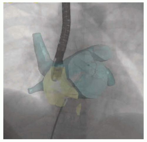
CT and MRI 3-D images are obtained prior to the procedure, and the images are processed and segmented to include the anatomical area of interest. The data are then transferred to a workstation in the catheterization lab, where the images are scaled and overlaid on the fluoroscopy image in 3-D space using anatomical landmarks such as the vertebral bodies or cardiac borders. This allows the interventional cardiologist to maintain alignment of the two images when the fluoroscopy detector is rotated. The 3-D image will maintain the alignment and project a 3-D image of cardiac structures and soft tissues (Fig. 38-1) (9). This can help reduce fluoroscopy time and contrast usage in some cases.

CT imaging and angiography have proven to be of critical importance in the evaluation and planning of certain structural cardiac interventions. Different imaging protocols to evaluate the cardiac as well as the vascular bed and the aorta are crucial in some cases to decide on the route of access and plan for the cardiac intervention. CT systems with at least 64-detector technology are recommended. This will give sufficient spatial resolution of cardiac and vascular structures; however, the temporal resolution remains inferior to that of echocardiography. Some of the disadvantages of CT imaging are related to the use of iodinated contrast and the exposure to ionizing radiation. Using electrocardiography (ECG) synchronization, advanced scanner technology and imaging protocols can reduce the radiation exposure significantly (Table 38-3) (10).

Cardiovascular MRI has the advantage of visualizing the cardiac and vascular structures without exogenous contrast. Other advantages of MRI include high spatial resolution and excellent soft tissue characterization. The main drawback of using RT MRI for catheter-based interventions is due to the compatibility of procedural hardware and catheters with the magnet and the limitation due to respiratory and cardiac motion with the need for rapid image reconstruction and display. The same mechanism that provides unique image quality also makes polymer-only catheters invisible and metal braided catheters to cause artifact and obscure an entire organ. Most of the equipment used in the catheterization laboratory is not MRI-safe, and hybrid MRI/Fluoroscopy suites require dedicated equipment, trained personnel, and additional space and cost. RT MRI is available in hybrid suites but is being used mainly for investigational therapeutic procedures (11).


Get Clinical Tree app for offline access