Patients with hepatic hemangiomas have been known to have high-output heart failure as a result of left-to-right arteriovenous shunting. We report a patient with a hepatic hemangioma that presented with high-output heart failure with hypoxia on exertion. After embolization of the hemangioma, the patient’s hypoxia resolved and ejection fraction improved. In the absence of cardiopulmonary pathophysiology, we presume that our patient’s hemangioma was causing a right-to-left shunt as opposed to an expected left-to-right shunt.
Hemangiomas have been known to induce high-output heart failure as a result of left-to-right systemic shunting. There is no known report of a hemangioma causing right-to-left systemic shunting with associated hypoxia. We present a case of high-output heart failure with exertion-induced hypoxia from a hepatic hemangioma with resolution after embolization.
Case Description
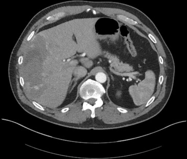
The patient is a 57-year-old white man with a history of well-controlled hypertension who was referred for dyspnea and neck tightness on exertion. Chest radiography and pulmonary function tests with spirometry were unremarkable. Electrocardiogram showed normal sinus rhythm with a left axis deviation and intermittent premature ventricular complexes. The hemoglobin was 16.5 g/dl, a brain natriuretic peptide was 18 ng/L, and a d -dimer was <0.19 μg/ml. Serum chemistry was unremarkable with sodium of 141 meq/L, potassium of 4.0 meq/L, albumin of 4.2 g/dl, alanine transferase of 26 U/L, aspartate transferase of 44 U/L, total bilirubin of 0.83 mg/dl, and creatinine of 0.9 mg/dl. Exercise stress testing (9 minutes 26 seconds on a Bruce protocol) showed >1 mm of ST depression in leads II and aVF and a peak stress pulse oximetry of 82%. Oxygen saturation at rest was 92%. Transthoracic echocardiography revealed an ejection fraction of 45% with mild global hypokinesia. Computed tomography of the chest exposed an incidental finding suggestive of a large neoplasm of the right hepatic lobe with no evidence of parenchymal lung disease. Computed tomography of the abdomen ( Figure 1 ) revealed a 14 × 6-cm right hepatic lobe mass with nodular peripheral arterial enhancement, gradually filling in on venous phases, consistent with a hemangioma. Coronary angiography showed normal coronary arteries with right dominant circulation. Cardiac catheterization data are listed in Table 1 . Hepatic arteriography ( Figure 2 ) confirmed the computed tomography findings and hepatic transarterial embolization was performed using 350- to 500-μm polyvinyl alcohol foam particles. Three months later, a follow-up exercise stress test (10 minutes on a Bruce protocol) showed no ischemic changes on electrocardiogram with a peak stress O 2 of 94%. Follow-up echocardiography revealed an improved ejection fraction of 55%.
| Heart Rate (beats/minute) | 60 |
| Aortic Pressure (mmHg) (S/D) | 128/78 |
| LV Pressure (mmHg) (S/D) | 119/1 |
| LV EDP (mmHg) | 4 |
| Arterial O 2 (%) | 92 |
| RA Pressure (mmHg) (S/D) | 3 |
| RV Pressure (mmHg) (S/D) | 31/1 |
| RV EDP (mmHg) | 1 |
| PA Pressure (mmHg) (S/D) | 26/8 |
| PWP (mmHg) | 3 |
| Cardiac Output (L/min) | 9.46 |
| Cardiac Output Index (L/min/m 2 ) | 4.08 |
| Middle RA O 2 (%) | 75 |
| Right Ventricle O 2 (%) | 75.6 |
| PA O 2 (%) | 77.9 |
| Superior Vena Cava O 2 (%) | 74.6 |
| Inferior Vena Cava O 2 (%) | 73.1 |
| Pulmonary Vascular Resistance (WU) | 1 |
| Systemic Vascular Resistance (WU) | 10 |
Stay updated, free articles. Join our Telegram channel

Full access? Get Clinical Tree


