Heart Transplantation
Heart transplantation has emerged as an effective therapy for patients with end-stage heart diseases. In 2005, a total of 2,125 heart transplants were performed in the United States. The major obstacle to more widespread application of heart transplantation is donor shortage.
Donor Selection
Matching of a donor heart to a specific recipient requires consideration of many donor and recipient factors, several of which have changed over time. Although there is no absolute maximum age for cardiac donors, many centers use an upper age limit of 55 to 65 years.
A history of diabetes mellitus in the donor with microvascular disease, long-standing donor hypertension with left ventricular hypertrophy (by electrocardiogram or echocardiogram), or prolonged high-dose donor heart inotropic requirement may be associated with an increased risk of early graft failure. Segmental or global wall motion abnormality of the donor heart can be associated with brain death, and should not be considered a contraindication to transplantation. Resuscitation with thyroid hormone or the addition of inotropes and/or vasoconstrictors may lead to improvement in left ventricular function. The donor can then be reassessed with a repeat echocardiogram or a pulmonary artery catheter.
Size matching of the donor and recipient is important. Severe undersizing can lead to the inability of the donor heart to support the recipient’s circulation, especially if there is evidence of primary graft dysfunction. Most programs require a donor to recipient weight ratio of at least 0.7. Oversizing can lead to restrictive physiology due to limited recipient mediastinal space. This issue is especially relevant in patients whose native heart disease is not dilated. Donor/recipient size matching has to be considered in association with other donor and recipient variables (i.e., an undersized female donor heart may not be suitable for a male recipient with pulmonary hypertension, especially in a setting of mild donor left ventricular hypertrophy and/or long ischemic time).
It is generally recommended that male donors older than 40 years and female donors older than 45 years undergo a coronary angiogram if available. Presence of significant coronary artery disease (> 50% lesions) in two or more major coronary arteries is usually a contraindication to utilization of a donor heart. However for critically ill recipients, donor hearts with discrete coronary stenoses can undergo bypass grafting using recipient conduits ex vivo, and be transplanted with acceptable short-term outcomes.
Aside from the considerations mentioned earlier, other contraindications to the use of a donor heart include positive human immunodeficiency virus (HIV) serology, positive hepatitis C serology, donor malignancies other than primary brain tumor, and systemic bacterial infection (especially with gram-negative organisms).
Preservation Solution
The ideal preservation solution will ensure microvascular, cellular, and functional integrity of the donor heart during the ischemic phase. Experience with the currently used preservations solutions (University of Wisconsin and Celsior solution) have shown excellent myocardial functional recovery, especially when the ischemic time is less than 6 hours.
University of Wisconsin solution is an “intracellular” based solution (low sodium, high potassium) and contains several classes of impermeable molecules to minimize cellular swelling. Because of the concern about the deleterious effects of high potassium concentrations on microvasculature, Celsior solution, which is an “extracellular” solution, was developed. In addition to many impermeable molecules, Celsior also has glutamate that serves as a substrate for energy production. Several studies have shown that both solutions afford similar protection to the donor heart during preservation. We currently use University of Wisconsin solution as our preservation solution of choice.
Donor Operation
Upon arrival at the donor hospital, the procurement surgeon will review the donor’s medical records to ensure
the accuracy and completeness of all data. The donor is placed in supine position with arms extended by the side. Because most donors are multiorgan donors, the donor is prepped from neck to midthigh. Midline sternotomy incision is performed as previously described. In smaller community hospitals, a sternal saw may not be available and a Lebsche knife may be used. The pericardium is opened and pericardial sutures are placed. The right pleural space is opened widely. The heart is systematically examined for size, evidence of right ventricular dysfunction, contusion, aneurysm, segmental wall motion abnormality, or a thrill suggestive of valvular heart disease. The course of the coronary arteries is palpated for evidence of calcification or plaques. If the quality of the donor heart is acceptable, this information is communicated to the recipient hospital.
the accuracy and completeness of all data. The donor is placed in supine position with arms extended by the side. Because most donors are multiorgan donors, the donor is prepped from neck to midthigh. Midline sternotomy incision is performed as previously described. In smaller community hospitals, a sternal saw may not be available and a Lebsche knife may be used. The pericardium is opened and pericardial sutures are placed. The right pleural space is opened widely. The heart is systematically examined for size, evidence of right ventricular dysfunction, contusion, aneurysm, segmental wall motion abnormality, or a thrill suggestive of valvular heart disease. The course of the coronary arteries is palpated for evidence of calcification or plaques. If the quality of the donor heart is acceptable, this information is communicated to the recipient hospital.
The dissection of the donor heart is started by freeing the superior vena cava from pericardial reflection to the innominate vein. The azygous vein is usually tied and divided to ensure sufficient length of the superior vena cava.
 For recipients with congenital heart disease who have previously undergone a classic or bidirectional Glenn procedure, a longer segment of innominate vein may be required.
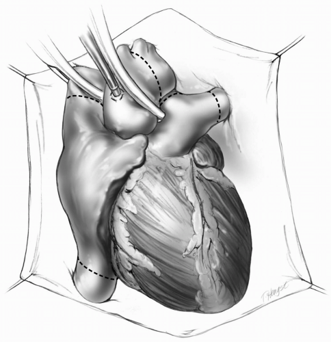
The aorta is dissected distally beyond the innominate artery take-off. The needle for administration of preservation solution is inserted into the ascending aorta and secured (Fig. 11-1). When the other procurement teams have completed their respective organ dissections, heparin at a dose of 300 units per kilogram of body weight is administered.
The most important step in heart procurement is to ensure that the donor heart is emptied. The pericardium on the right side is incised at the level of the hemidiaphragm down to the inferior vena cava. The superior vena cava is clamped and the inferior vena cava is transected so that the blood from the heart empties into the right chest cavity.
 If the lungs are being harvested, exsanguination has to be done into the abdomen by the abdominal team.
When the heart is empty (usually after 5 to 10 beats), the aortic cross-clamp is applied and the preservation solution is administered into the aortic root. We measure pressure in the ascending aorta and maintain it between 50 and 70 mm Hg. The apex of the heart is elevated toward the right side, and the left inferior pulmonary vein is incised where it joins the left atrium (Fig. 11-2). The pericardium is filled with ice slush to ensure topical cooling. A total of 10 mL per kg of donor body weight of University of Wisconsin solution is administered, which may take several minutes. During this time, the procurement surgeon must ensure that the heart is not distended by frequent palpation of the left ventricle. The donor heart usually stops beating after 30 seconds of perfusion with the preservation solution.
 FIG 11-1. The donor heart is prepared. Antegrade cardioplegia needle has been placed, and the aortic cross-clamp is applied. |
 When the lungs are also being harvested, the incision is made halfway between the left inferior pulmonary vein entry into the left atrium and the atrioventricular groove. This maintains adequate cuffs of pulmonary veins for lung harvest.
Stay updated, free articles. Join our Telegram channel

Full access? Get Clinical Tree


