Hamartoma
Presentation
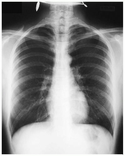
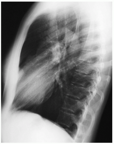
A 32-year-old woman who recently immigrated to the United States from Europe received a chest x-ray for the immigration process. She is a nonsmoker and worked as a kindergarten teacher in her homeland. She has no pain and denies cough. There is no family history of cancer. Vital signs are normal. Physical examination is unremarkable.
Differential Diagnosis
The differential diagnosis includes a number of benign, inflammatory, and malignant conditions. Hamartoma is the most common benign lung tumor, composing 5% of resected nodules. Other benign conditions of the lung include clear cell tumor, chondroma, fibroma, fibromyxoma, sclerosing hemangioma, and granular cell myoblastoma. Inflammatory nodules are more common than benign neoplastic tumors. Most are granulomas that result from prior infection of tuberculosis or fungi, such as Histoplasma, Coccidioides, or Blastomycosis species. Other causes of granulomas that can be multiple or solitary are sarcoidosis, Wegener’s granulomatosis, or eosinophilic granuloma.
Recommendation
If available, previous chest x-rays should be obtained for comparison; in addition, computed tomography (CT) scans are indicated to localize the nodule and plan a diagnostic and therapeutic approach.


