Extremity Causalgia: Reflex Sympathetic Dystrophy


Extremity Causalgia


Reflex Sympathetic Dystrophy


Kelley Hodgkiss-Harlow and Dennis F. Bandyk


Understanding the pathophysiology and management of extremity causalgia, a complex regional pain syndrome (CPRS), is relevant to the practice of vascular surgery. Patient referrals for extremity complaints (pain, cyanosis, skin temperature changes, edema) can mimic arterial and venous disease, but once a vascular abnormality is excluded, a neurogenic basis for the symptom complex should be considered.


Reflex sympathetic dystrophy (RSD), a type I CRPS, displays specific clinical features of burning extremity pain, sympathetic hyperactivity, muscle wasting, joint stiffness, and trophic skin changes. The condition is often underdiagnosed or misdiagnosed following trauma, with a prevalence of 2% to 5% following peripheral nerve injury, 1% to 2% after bone fracture, and less than 1% after soft tissue contusion or surgical procedures.


In 1995, the International Association for the Study of Pain recommended CRPS be classified into two types: type I or RSD, and type II, a pain syndrome with documented peripheral injury as the initiating factor. Although the CPRS classification has been adopted by pain specialists, the condition of RSD remains rooted in diagnostic terminology with patients and primary care physicians and in the determination of medical disability caused by workplace accidents. The term mimocausalgia has also been used to denote RSD symptoms occurring late after minor extremity trauma and should be considered type I CPRS, but the diagnosis of mimocausalgia can delay recognition of RSD and medical treatment owing to its uncommon usage.


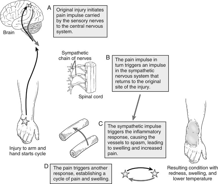
The development of RSD is poorly understood, but it is related to sympathetic nerve dysfunction and involves three nervous system mechanisms (Figure 1):


The onset of RSD following extremity trauma is variable, but it typically develops within weeks of the injurious event. Initially, the extremity pain and disability may be characterized by intervals of exacerbation followed by partial remission, but the normal healing process with pain resolution does not occur. Rather, chronic sympathetic nerve dysfunction triggers an inflammatory response producing cyclic vasospasm and pain sensitivity to even the slightest touch (tactile allodynia).


The pathophysiology of RSD results in the signs of mottled skin, muscle spasms, and joint immobility, but the primary compliant is a disabling burning pain in the extremity. This CPRS condition can develop in children and adults and has no apparent no gender predisposition. Its development is associated with a variety of events, with the common denominator being extremity injury (Box 1). Young (15-30 yr), active adults appear to be the most prone to RSD, which develops when extremity injury does not follow the normal path of healing. Development does not depend on the magnitude of injury, and initial diagnosis can be hampered by the lack of physical findings in the setting of severe extremity pain. Joint sprains and repetitive motion disorders, such as carpal tunnel syndrome, can produce RSD and result in accusations of malingering if the inciting event was work related.


There is no definitive radiologic or diagnostic test to confirm the diagnosis of RSD. Standard extremity x-rays can rule out bone disease, although chronic CPRS is associated with patch osteoporosis, which can be confirmed by a three-phase bone scan. Thermography indicates a cool affected extremity relative to the other limbs, and other signs of sympathetic hyperactivity (sweat output, skin color changes) are used to augment the clinical impression.


In 1959, Drucker and colleagues characterized three clinical stages of RSD with the progression of symptoms and disability occurring in an unpredictable manner with time (Box 2). In stage I RSD, extremity pain is localized to the region of injury but with pain increasing during the healing process. Extremity tenderness and sensitivity to normal touch or motion is out of proportion to what is expected based on objective signs (contusion, fracture, surgical site healing). The pain is described as constant, with features of a burning sensation or a deep ache. Any repetitive tactile contact escalates pain severity (allodynia) and causes the pain to persist for an extended time period (hyperpathia). Palpation of skin trigger points as seen with other myofascial pain syndromes can often be elucidated on questioning the patient or performing physical examination. Evaluating the patient by sympathetic block is both diagnostic and therapeutic, because the clinical course of acute RSD may be reversible, and regional sympathetic block can provide pain relief well beyond the normal duration of the local anesthetic drug used.


Progression of RSD to stage II is manifest by the development of visible skin changes, including cold sensitivity, cyanosis, sweating or skin dryness, muscle atrophy, and joint stiffness. Cold sensitivity is present with all RSD stages and typically is associated with manifestations of excessive sympathetic tone such as hyperhidrosis, cool skin temperature, and pilomotor changes. Abnormalities of bone structure (osteoporosis), hair growth, and nail texture (brittleness) can occur. This dystrophic stage of RSD is associated with a positive bone scan demonstrating increased calcium absorption. Limb swelling is also a common sign of RSD progression and can develop into a hard, brawny form of lymphedema, especially in the lower extremity. Muscle and joint stiffness produces decreased range of motion and may be accompanied by involuntary spasms and eventually limb disuse. Pain relief with a sympathetic block indicates a sympathetic-maintained pain syndrome, but the condition is not unlikely to spontaneously resolve without proper therapeutic intervention.


In stage III RSD, the condition is associated with extremity atrophy and intractable pain. The likelihood that pain relief occurs with sympathetic block decreases, and if a block produces no pain relief, the condition is termed sympathetic independent. The burning pain can spread beyond the affected extremity to the trunk, face, and other extremities (upper or lower). The onset of spreading RSD can occur without an identifiable traumatic event. If symptoms are associated with excessive sympathetic activity, sympathetic blocks and sympathectomy might provide pain relief to the other involved extremities.


Early diagnosis of RSD coupled with medical and rehabilitative therapy, including sympathetic nerve block therapy, can be curative. Even when RSD symptoms resolve, the extremity remains at risk for recurrence months to years later. Minor traumatic events can reactivate the RSD condition. In the majority of patients with symptoms lasting months, the RSD syndrome develops into a chronic and permanent disability affecting daily activities, ability to work, and social relationships.


Differential Diagnosis


The diagnosis of RSD is often delayed as a result of its varied clinical presentation and lack of a definitive test. The hallmark of CPRS is extremity pain and mobility problems out of proportion to those expected from the original injury. The arterial and venous examination is normal, including normal limb and digit systolic blood pressure measurements and normal Doppler phasic artery and venous flow signals. An important diagnosis to consider, especially following a surgical procedure, is nerve entrapment. Causalgia pain can develop if a nerve is injured, trapped in the surgical site closure, or encased by developing scar tissue. Often, focal palpation at the site of nerve injury or entrapment will elicit a sharp radiating pain in the nerve’s sensory distribution. Infiltration of a local anesthetic into the region relieves the pain and confirms the diagnosis.


The clinical features of acute RSD with cyclic symptom exacerbation requires an astute physician to consider the diagnosis and refer to a multidisciplinary pain clinic. A multidisciplinary approach to the CPRS patient is recommended to exclude other musculoskeletal and peripheral nerve conditions that contribute to the extremity pain, swelling, and disability. In the presence of edema, the extremity should be assessed by venous duplex ultrasound to assess the patency and valve function of both the superficial and deep veins. The presence of soft tissue edema, hematoma, or joint cysts may be identified by B-mode imaging and help in establishing a diagnosis. Careful examination of the musculoskeletal system is mandatory, including presence of joint space effusion, range of motion, and muscle strength and function. Assessment for muscle atrophy by visual inspection and measurement of limb girth should be performed and compared with the contralateral unaffected extremity. Documentation of skin color, trophic changes, and signs and symptoms of excessive sympathetic activity, including during stressful conditions, should be performed. The neurologic examination should focus on both motor and sensory nerve function and on whether an abnormal sensory response occurs with tactile stimulation.


The nature and severity of the extremity pain should be determined using a visual analogue pain severity scale. This assessment is useful to establish baseline pain severity and document changes at each subsequent patient evaluation. The patient grades the basal pain severity on a visual analogue scale from 0 to 10, where 0 is no pain, 5 is pain that interferes with daily activities, and 10 is the worst pain imaginable. The typical RSD patient rates pain severity in the range of 7 to 8, with daily occurrence of exacerbations to 10. This level of pain interferes with all social relationships, it results in inability to work, and the patient is commonly housebound.


An important test to establish the diagnosis of RSD is sympathetic blockade via the stellate ganglion (upper extremity symptoms) or lumbar ganglion chain (lower extremity symptoms) by infiltrating a local anesthetic (lidocaine, bupivacaine). Selective sympathetic nervous system block provides useful information as to whether the pain is sympathetically maintained and is therefore potentially responsive to further temporary blocks, chemical sympathectomy, or surgical sympathectomy. Patients are instructed to quantitate the degree of pain relief obtained by the sympathetic block as a percentage of their basal pain severity, for example, 100% relief, 50% relief, or no relief. The degree of pain relief is an excellent predictor of pain relief that can be expected with chemical or surgical sympathectomy. A greater than 50% decrease in pain severity lasting longer than 2 days following a sympathetic block indicates a sympathetic-maintained pain syndrome. In such patients, a series of three to six sympathetic blocks can produce a cure or partial RSD remission.

Stay updated, free articles. Join our Telegram channel

Aug 25, 2016 | Posted by in CARDIOLOGY | Comments Off on Extremity Causalgia: Reflex Sympathetic Dystrophy

Full access? Get Clinical Tree

Get Clinical Tree app for offline access