Esophageal Adenocarcinoma and Transhiatal Esophagectomy
Presentation
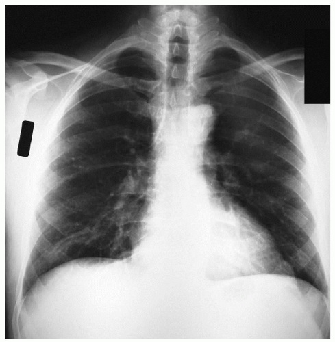
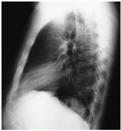
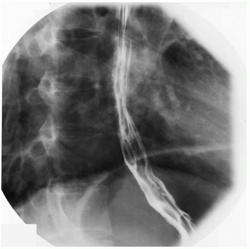
A 51-year-old man is admitted to the emergency department with a 2-month history of worsening episodes of dysphagia. He complains of retrosternal discomfort soon after ingesting solid food that lasts about 10 minutes. He has lost 10 pounds in the past 2 months. He is a nonsmoker but drinks one or two bottles of beer per day. On physical examination, the patient is somewhat cachectic, and his vital signs are stable. He has the following admission chest x-rays and esophagogram.
▪ Chest X-rays
Chest X-ray Report
There are no lung masses. The heart is normal in size. There are no pleural effusions. ▪
Case Continued
The patient is discharged from the emergency department with instructions to follow up with his primary physician. After several weeks, the patient continues to complain of dysphagia. Based on the clinical symptoms, his physician suspects a motility abnormality and refers the patient to a gastroenterologist for an esophagoscopic study.
▪ Esophagoscopies
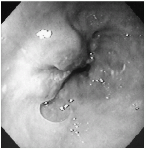 Figure 34-4 See Color Plate 15 on page 114. |
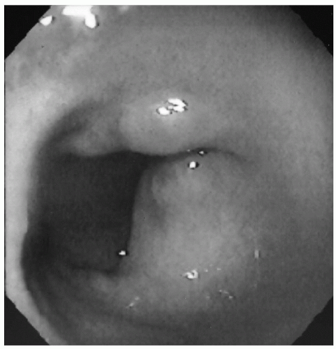 Figure 34-5 See Color Plate 16 on page 114. |
Esophagoscopy Results
Stay updated, free articles. Join our Telegram channel

Full access? Get Clinical Tree


