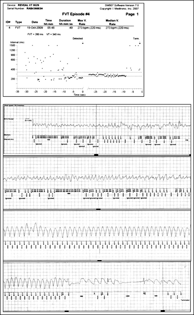
The images of figure 1 and 2 were switched, i.e., in the published paper the image in figure 1 corresponds to figure legend 2 (VF initiated by early coupling VPC, as recorded on telemetry during hospitalization.), whereas, the image in figure 2 corresponds to figure legend 1 (ILR tracing demonstrating VF that organized into monomorphic VT, followed by spontaneous termination and sinus bradycardia). The correct order is shown on the following two pages:


