Endovascular Treatment of Nonruptured Infrarenal Aortic and Aortoiliac Aneurysms


Endovascular Treatment of Nonruptured Infrarenal Aortic and Aortoiliac Aneurysms


Gavin C. O’Brien, Raffi A. Qasabian, and Geoffrey H. White


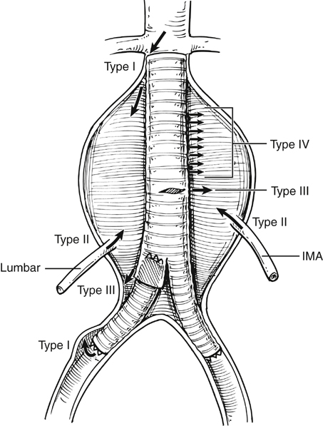
Endovascular aneurysm repair (EVAR) has become established as the first-line approach to treating abdominal aortic aneurysms (AAAs). Conceptually, the idea of using vascular endoprosthesis to exclude aneurysms dates back to the late 1960s with animal experimentation. The landmark first deployment of an aortic stent to exclude a human AAA was reported by Parodi and colleagues in 1991. Initially, straight grafts were used, with polyester tubes being held in position within the aorta by large Palmaz stents. It was soon realized that as few as 5% of infrarenal AAAs had an anatomy suitable for such a device, resulting in a high incidence of failure as a result of type I endoleaks (Figure 1) With time, modular bifurcated grafts became the preferred option for infrarenal AAA repair.


Procedural Planning and Anatomic Measurements


Preprocedural planning is one of the most important aspects required for achieving success with endovascular AAA repair. The major anatomic regions of interest in the planning process are the aortic neck and the iliac arteries. Problem features in the neck include angulation, irregular shape, short length, and poor aortic wall quality. Difficult necks are a significant cause of procedure failure modes such as device migration, endoleak, and late aneurysm rupture. Iliac artery anatomy also has significant influence, with angulated or calcified arteries being risk factors for failed access, iliac rupture, and device thrombosis.


Procedural planning for EVAR operations starts with a careful assessment of the imaging studies. The essentials of the aortoiliac and branch anatomy should be measured by electronic digital image calibration and then recorded on a dedicated planning sheet. These aspects include the basic dimensional details of the aortic neck (neck diameter, length, shape) as well as other characteristics such as angulation in the anteroposterior or lateral planes, wall irregularity, and existence of thrombus. Also recorded are the shape and diameter of the flow lumen through the sac and the characteristics of the access arteries (iliac tortuosity, calcification, and atheroma). It is important to ascertain the number of renal arteries and their relationship to the aortic neck, as well as the position of the lowest renal artery with respect to bony landmarks, such as the first or second lumbar vertebrae, and any significant amount of thrombus or atheroma involving the aortic wall close to the renal orifices or within the aortic neck.


This information is used to plan the device dimensions and the order of the procedure, including a consideration of likely difficulties or complications during the operation and a backup plan for their management. Planning should also include the provision of reserve or extension grafts, which can be used to deal with endoleaks or inadequate coverage. This is particularly applicable in patients with a difficult aortic neck or calcified, atherosclerotic iliac arteries.


Measurements of the diameter of the aortic neck and the iliac arteries are, in general, best obtained from computed tomography (CT) scan images, whereas length measurements are more accurate when obtained from three-dimensional (3-D) reconstructions or from calibrated angiography. When measuring vessel diameters, it is important to recognize that device companies can differ in requesting internal or external (adventitial) diameters. Vessel diameter measurements usually assume a circular or cylindrical shape, so it is useful to verify that the actual shape is not ovoid or irregular by referring to the 3-D images. Most interventionists now use reformatting programs and specialized workstations such as the TeraRecon or Osirix to create 3-D images to adequately plan procedures. Very occasionally, adjunctive calibrated angiography can help with procedure planning, particularly in the presence of a short or severely angulated neck, especially if the neck is angulated in the anteroposterior plane. The angles of deployed devices should be anticipated, and consideration should be given to whether this could be improved by using a different access side or technique or by using different device components.


This information transposes into intraprocedural imaging, with appropriate planning and orientation of the image intensifier in anteroposterior or lateral planes. This should reduce the hazards of parallax error in deploying the device, particularly in the setting of an angulated aortic neck or tortuous iliac arteries, thus reducing the risk of covering the renal or internal iliac arteries. Extra graft extensions of various sizes, balloons, access catheters, and guidewires should be available. In cases where there access may be difficult, an alternative technique of access should be preplanned: for example, iliac conduit graft by using a retroperitoneal approach.


Device Oversizing


Most endovascular device companies recommend use of a graft that is oversized by approximately 10% to 15% compared to attachment zone diameters in the aortic neck and the iliac arteries, to attain both attachment and seal within these sites. Usually the aortic neck diameter is calculated with respect to the external (adventitial) surface, so that the true diameter of the lumen may be considerably less and the graft diameter may therefore be oversized substantially more with respect to the lumen.


Excessive oversizing (20% or more) can cause the neck to dilate owing to the excessive radial expansile force and has been shown to be associated with increased graft migration rates with at least two self-expanding device designs. In one study, oversizing by more than 20% with the AneuRx graft resulted in late aortic neck dilation and high rates of device migration. With the Zenith graft, oversizing of 30% in a multicenter phase II trial resulted in a 14-fold increase in migration rates, as well as increased rate of AAA sac enlargement and early dilatation of the aortic neck at the 6-month follow-up interval, which then stabilized at up to 24 months. Although these changes may be device dependent, avoidance of excessive oversizing is recommended. The characteristics of the neck itself are also important factors: A short neck or aortic neck angulation greater than 40% can have significant adverse effects on the outcome.


Endovascular Stent Deployment Techniques


Imaging


Procedures should ideally be performed in an environment that has the capacity for both surgical exposure of vessels (and the possibility of unexpected conversion to an open procedure) as well as excellent vascular imaging. Traditionally, EVAR was either performed in a standard surgical theatre suite with a mobile C-arm image intensifier and radiolucent sliding tabletop or in a modified radiology suite with enhanced surgical equipment. Currently, the emergence of a fully equipped hybrid suite combines these elements in a stand-alone dedicated endovascular operating theater.


Access


For many years, an open surgical approach to the femoral arteries was used, with an oblique transverse incision preferred by many owing to the perceived lower incidence of postoperative seroma or lymphocele compared to that when using a vertical incision. The incision is limited to 3 to 4 cm in length, because exposure of only the common femoral artery is required. Most access sheaths are designed with a smooth taper so that the sheath can be inserted directly over the guidewire into the artery, resulting in a gradual dilatation of the arteriotomy. Formal open surgical arteriotomy is limited to selected vessels that are severely calcified or stenotic. In the modern era, access to the femoral arteries is predominantly achieved using a percutaneous approach rather than by open femoral cutdown access.


Percutaneous closure devices such as the ProGlide device are deployed in the initial operative phase. The percutaneous approach appears safe and effective in systematic reviews, with success rates of 92% and complication rates of 4.4% reported for percutaneous access. These devices received investigational device exemption (IDE) conditional approval from the U.S. Food and Drug Administration in 2010 to begin a prospective multicenter randomized clinical trial for a bilateral percutaneous approach to EVAR (PEVAR trial). In Australia, more than 70% of EVARs are currently performed percutaneously. One would expect the complication rates of percutaneous closure to reduce further with the current development of lower-profile delivery systems.

Stay updated, free articles. Join our Telegram channel

Aug 25, 2016 | Posted by in CARDIOLOGY | Comments Off on Endovascular Treatment of Nonruptured Infrarenal Aortic and Aortoiliac Aneurysms

Full access? Get Clinical Tree

Get Clinical Tree app for offline access