Endovascular Angioplasty and Stenting for Proximal Subclavian Artery Stenosis


Endovascular Angioplasty and Stenting for Proximal Subclavian Artery Stenosis


Ali F. AbuRahma and Patrick A. Stone


The prevalence of atherosclerotic stenotic disease involving the proximal brachiocephalic arteries is significantly less than that in the extracranial carotid arteries. Additionally, only 10% of patients with hemodynamically significant proximal subclavian artery stenoses develop symptoms. This probably reflects the robust collateral network distal to ostial subclavian artery stenosis, including the vertebral and internal mammary arteries. Unfortunately, there is a paucity of data on the natural history of untreated proximal subclavian artery stenosis.


The incidence of subclavian steal syndrome was evaluated in more than 7881 patients undergoing carotid artery duplex scanning. A pressure difference of more than 20 mm Hg, indicating subclavian artery stenosis, was found in 514 (6.5%); however, symptoms were only present in 38 patients, with the majority being related to the posterior circulation. Patients with a difference of greater than 40 mm Hg in the extremities were more likely to have associated symptoms. In patients undergoing evaluation for coronary artery disease, 2.5% were found to have significant proximal left subclavian artery stenosis. The MESA study (Multi-Ethnic Study of Atherosclerosis) evaluated 6743 patients and reported that 4.6% of patients had subclavian stenosis, as defined by a systolic blood pressure difference of 15 mm Hg. There was also a higher prevalence in women (5.1%) compared with African Americans and men (3.9%).


Clinical Evaluation and Diagnosis


Most patients with subclavian stenosis are asymptomatic. Patients are often referred for evaluation by primary care physicians after they find asymmetry in the upper extremity blood pressures. A brachial blood pressure difference exceeding 15 to 20 mm Hg is considered a significant finding.


Symptoms of subclavian artery stenosis can occur either in the form of exertional ischemia of the extremity (limb fatigue or pain) or, rarely, digital embolization, which can manifest as ulcerations or nonhealing wounds. Vertebrobasilar symptoms can occur in patients with reversal of flow in the vertebral artery, and this is referred to as subclavian steal syndrome. Angina or acute myocardial infarction can occur in patients with a previously placed internal mammary bypass and hemodynamically significant proximal subclavian stenosis.


Rarely, patients present with pseudohypotension and are found to have severe bilateral subclavian artery stenoses. Physical examination should include an inspection of the hand and digits for evidence of embolic phenomena; palpation of the brachial, radial, and ulnar arteries; auscultation of the supraclavicular fossa for bruits; and a recording of bilateral brachial artery pressures.


Duplex examination should be the first imaging modality to assess patients with suspected symptomatic disease. Standard imaging of the extracranial carotid arteries should be performed to assess for concomitant disease affecting these vessels, as well as an indirect assessment of the proximal carotid arteries by waveform and velocity analysis. A duplex examination of the subclavian and axillary arteries can suggest significant proximal subclavian artery disease in the presence of elevated peak systolic velocities or monophasic waveforms. However, no recognized duplex criteria have been widely accepted in assessing these arteries. A duplex examination of the vertebral artery should include direction of blood flow: antegrade, bidirectional, or reversed. Bidirectional or reversed flow suggests high-grade subclavian stenosis or occlusion or disease of the innominate artery.


Conventional subtraction angiography is the gold standard for establishing the diagnosis of a subclavian arterial stenosis and has the added benefit of an optional therapeutic intervention during the diagnostic procedure. Computed tomography and magnetic resonance arteriography have limited utility for evaluating subclavian artery stenosis before conventional angiography.


Indications for Treatment


We consider asymptomatic, good-risk patients with severe bilateral subclavian artery disease for treatment in order to facilitate accurate ambulatory blood pressure measurements. Additionally, asymptomatic patients with severe proximal left subclavian artery stenosis should be considered for intervention before coronary artery bypass grafting if the left internal mammary artery is to be used as a conduit. Symptomatic patients with exertional arm ischemia, vertebrobasilar symptoms such as subclavian steal, or angina related to a previous left internal mammary artery bypass are also offered treatment. Patients with vertebrobasilar symptoms or symptoms of subclavian steal with concomitant significant carotid artery stenosis should undergo carotid reconstruction prior to subclavian reconstruction, which may relieve their symptoms.


Endovascular treatment of subclavian artery stenosis can be performed with local sedation and minimal morbidity, but the threshold for intervention should not be lower than that of open surgical therapy. Additionally, although endovascular techniques have continued to improve since the turn of the century with lower-profile delivery systems, better stent design, and other advances, we do not agree that endovascular intervention is the first line of treatment in all patients or lesions. Nevertheless, most centers pursue angioplasty and stent placement, particularly in the management of subclavian and proximal carotid artery pathology.


Technical Principles of Endovascular Therapy


Arterial access for diagnostic procedures and interventions on the proximal subclavian artery may be obtained through the femoral, brachial, or radial arteries. The preferred approach is the femoral artery. Brachial access has been fraught with more access-related complications when compared with the femoral access. Others gaining access through the radial artery have reported excellent technical success with limited access complications. Once access is achieved, arch angiography at 30 degrees left anterior oblique is performed with a pigtail catheter.


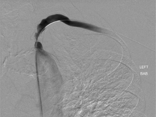
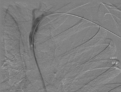
Contrast injections at 15 to 20 mL/second, for a total injection of 30 to 40 mL, are routine. Evaluation of the great vessels should include type of aortic arch, degree of aortic disease, and status of contralateral vertebral artery. The length and severity of subclavian artery stenosis should be assessed, along with the proximity of the lesion to the vertebral and internal mammary arteries (Figure 1). Selective catheterization can be performed with a variety of catheters, based on the preference of the interventionalist and the type of aortic arch.


Stenoses of greater than 75% are crossed with a hydrophilic wire after administration of 3000 to 5000 IU of unfractionated heparin. Purchase of the hydrophilic wire is achieved in the brachial artery. The diagnostic catheter is advanced and exchanged over a stiff wire; our preference is a 0.35-inch, 300-cm Hi Torque SupraCore Wire (Abbott Vascular, Calif.). The short 5-Fr sheath can be exchanged over the stiff wire for a long 6-Fr sheath in preparation for an intervention.


Critical ostial stenosis should be predilated to allow safe advancement of a balloon-expandable stent across the lesion. The sheath should then be advanced just distal to the lesion (Figure 2). Ostial lesions are best treated with balloon-expandable stents and should be sized 1:1 with the native artery. Oversizing in this setting can result in arterial perforation. If the surgeon is unsure of the size of the native artery, intravascular ultrasound can be used to obtain a measurement. After the balloon-expandable stent is advanced to the end of the sheath, the sheath is withdrawn into the aortic arch, leaving the stent across the lesion. Only 2 mm of the proximal stent should protrude within the arch of the aorta. If the protrusion of the proximal stent is too long, this can limit future repeat interventions and can pose a hazard with future coronary catheterizations. Currently, there is no Federal Drug Administration–approved stent for use specifically in the subclavian artery, and off-label use is required if primary stenting is chosen or needed if an inadequate angioplasty is achieved.


Stay updated, free articles. Join our Telegram channel

Aug 25, 2016 | Posted by in CARDIOLOGY | Comments Off on Endovascular Angioplasty and Stenting for Proximal Subclavian Artery Stenosis

Full access? Get Clinical Tree

Get Clinical Tree app for offline access