Emerging Technologies and Techniques for the Management of Non-Small-Cell Lung Cancer
Ricardo S. Santos
Fawad Khawaja
Hiran C. Fernando
Lung cancer is a significant public health problem, with approximately 215,000 new cases diagnosed annually in the United States.1 Lobectomy remains the mainstay of therapy for early stage non-small-cell lung cancer (NSCLC). Sublobar resection (SR) is generally regarded as a compromise operation, because of increased local recurrence, and is reserved for patients with impaired cardiopulmonary function.20 Approximately 15% to 20% of patients with stage I or II NSCLC are unable to undergo any operation owing to existing comorbid diseases.2 The standard therapy for these high-risk patients has been external beam radiation (EBR). Although the results with radiation have been better than with no treatment, treatment results with EBR are still inferior when compared with resection.21 In a study of 71 node-negative patients who received at least 60 Gy to their cancers, 3- and 5-year survivals were 19% and 12% respectively.15 Another study from Duke University of 141 stage I patients undergoing radiation therapy demonstrated 2- and 5-year survivals of 39% and 13% respectively.36
A concern with EBR is injury to surrounding tissues and the difficulties of delivering a precise area of radiation to a target area because of lung movement with respiration. Radiation pneumonitis can be life-threatening, particularly in patients with impaired pulmonary function. The rate of radiation pneumonitis has been reported to be around 8.3% with EBR.45
Over the last few years a number of new approaches for treating lung tumors in the high-risk patients have been introduced into clinical practice. Although large-scale studies and long-term results are not yet available, preliminary results are encouraging. This chapter reviews these approaches and their potential role in treating NSCLC. The approaches discussed include (a) SR with adjuvant brachytherapy, (b) radiotherapy ablation, and (c) stereotactic body radiation therapy (SBRT).
Sublobar Resection with Adjuvant Brachytherapy
The principal concern with sublobar resection (SR) has been the higher local recurrence that is seen compared with lobar resection. In a retrospective series by the Rush-Presbyterian group, a 22% local recurrence rate for patients with stage I NSCLC was reported after SR, compared with 4.9% after lobectomy.42 The Lung Cancer Study Group subsequently reported a randomized study of patients with T1N0 NSCLC undergoing lobectomy compared with SR.20 The principal finding in this study was a threefold increase in local recurrence in patients who had SR.
A number of factors may contribute to the consistent finding of increased local recurrence after SR. One issue may be that patients undergoing SR may be understaged if lymph node sampling or dissection is not performed. Nodal recurrence may simply represent progression rather than recurrent disease. A second factor may be that the margins of resection may be close or even positive, leading to increased parenchymal recurrence. A study by Goldstein et al.11 assessed T1N0 peripheral adenocarcinomas treated by wedge resection followed by lobectomy.11 Residual adenocarcinoma was found in 45% of the completion lobectomy specimens. The mean microscopic margin was 0.7 mm in these patients with residual disease compared with 2.4 mm in those without. Gross margins will frequently underestimate the microscopic margin. In the same study by Goldstein,11 the mean gross margin was 4.1 mm, compared with 2.3 mm for the mean microscopic margin. El-Sherif et al.8 recently reported results in 81 patients with stage T1N0 NSCLC who had undergone SR. Within this group there were 41 who had margins <1 cm and 40 with margins ≥1 cm. Local recurrence was 14.6% in patients with a margin <1 cm compared with 7.5% (p = 0.04) in patients with a margin >1cm.
Several groups have investigated different techniques to assess cytologic verification of margin negativity. Higashiyama and colleagues,13 using a lavage cytologic technique, demonstrated that although surgical margins were negative by frozen section, malignant cells were left behind. For those patients demonstrating positive cytological residua within the remaining pulmonary margins, local recurrence rates were higher. Sawabata et al.33 utilized a simpler technique to assess cytologic margins by brushing a slide along the staple line after performing SR. Their methodology revealed a 47% positive rate for residual malignant
cells, compared with only 20% with histologic techniques. Those patients with cytologically positive cells demonstrated a 57% margin relapse rate, compared with 0% amongst those with negative cytologic margins.
cells, compared with only 20% with histologic techniques. Those patients with cytologically positive cells demonstrated a 57% margin relapse rate, compared with 0% amongst those with negative cytologic margins.
When SR is utilized for patients with compromised pulmonary function, the operation can be challenging, and in many cases margins may be closer than desired. There will also be patients with histologically negative margins who will have cytologically positive margins as described above.
Adjuvant EBR is an option that has been reported to decrease local recurrence after wedge resection.25 In many ways this approach is analogous to the combination of lumpectomy and EBR, which has become standard therapy for many patients with anatomically favorable breast cancers. However, in the lung, difficulties in precisely delivering and developing a physics treatment plan for EBR to the staple line may occur because of physiologic lung motion and the three-dimensional irregularity of the target staple line following SR. SR and “postage stamp” EBR therapy was recently investigated in a study coordinated by the Cancer and Leukemia Group B.35 In their study, “extended radiotherapy fields were often necessitated by the extent of the postoperative staple line”; additionally, the logistic concerns of daily radiotherapy placed a hardship on some patients. Radiation pneumonitis is also a concern in these patients, even with postage-stamp radiation. An alternative to EBR is the use of intraoperative adjuvant brachytherapy. Two techniques have been described. The first involves the placement of paired sutures adjacent to the staple lines.17 These sutures contain iodine-125 (125I) seeds that deliver the radiation, effectively increasing the treated margin. The sutures are a commercially available product (Oncura, Arlington Heights, Illinois). The second technique involves placement of the same sutures into a Vicryl mesh that is then placed over the staple line4 (Figure 119-1). Typically about four parallel suture strands are placed in the mesh to deliver a dose of 10,000 to 12,000 cGy to the resection margin. The advantage of brachytherapy is that this provides a means of delivering radiation in a more uniform manner, with 100% patient compliance and in the same setting as the lung resection. One study17 reports the use of brachytherapy in 33 high-risk patients, primarily after wedge resection. The local recurrence rate was 6.1%, which was similar to the 6.4% reported after lobectomy in the Lung Cancer Study Group (LCSG) study.20 Another study from Pittsburgh32 compared local recurrence rates in 98 patients undergoing SR with brachytherapy with those of 102 patients undergoing SR alone. There was no difference in operative mortality, distal recurrence, or survival. However, local recurrence was significantly reduced from 18.6% to 2% in those patients who had adjuvant brachytherapy. It should be emphasized that this second study consisted primarily of segmental resections compared with the other study, which consisted of mostly wedge resections. In a more recent multicenter retrospective study looking at stage IA cancers, brachytherapy was used in 60 of 124 SR resections.10 Local recurrence was significantly lower in those patients who had brachytherapy at 3.3%, compared with 17.2% for those patients treated with SR alone. The results of these preliminary studies are encouraging and have led to a phase III study (Z4032) of this technique, which is currently being undertaken by the American College of Surgeons Oncology Group. It is hoped that this clinical trial will better define the role of brachytherapy following SR.
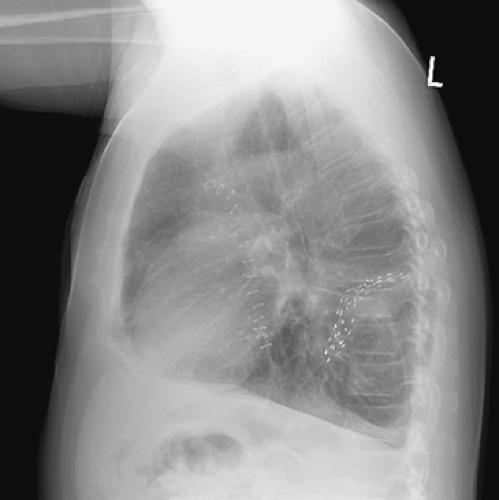 Figure 119-1. Chest x-ray demonstrating mesh implant after superior segmentectomy. |
Radiofrequency Ablation
Radiofrequency ablation (RFA) is a relatively new modality that has been successfully used for the treatment of hepatic tumors.3 Over the past few years there have been an increasing number of reports describing the use of RFA for malignant lung nodules.7,34 Most reports have demonstrated the feasibility of this technique; studies with intermediate follow-up are now being reported with increasing frequency.10,28,37
Radiofrequency (RF) energy consists of an alternating current that moves from an active electrode placed within a tumor to dispersive electrodes (electrosurgical return pads) placed on the patient. RFA systems therefore comprise three components: a generator, an active electrode, and dispersive electrodes. As the RF energy moves from the active electrode to the dispersive electrode and then back to the active electrode, ions within the tissue oscillate in an attempt to follow the change in direction of the alternating current. This results in frictional heating of the tissue; as the temperature within the tissue becomes elevated beyond 60°C, cells begin to die. It is this phenomenon that causes the region of necrosis surrounding the electrode.
The advantage of such a thermal intervention system is the capacity to heat tissue to a lethal temperature in a specific anatomic location. This allows for destruction of tumor tissue with minimal damage to surrounding normal tissue, which is particularly important in the patient with pulmonary compromise.
Currently three RFA systems are approved by the FDA for the ablation of soft tissue. These are the Boston Scientific (Natick, Massachusetts), RITA (RITA Medical Systems, Fremont, California), and Valleylab (Boulder, Colorado) systems. The Boston Scientific (BOS) and RITA probes contain multiple tines that are
expanded within the tumor nodule being ablated (Figs. 119-2 and 119-3). The Valleylab (VL) probe consists of either a single needle, or three parallel needles (cluster probe) placed into the tumor nodule (Figure 119-4). The VL probe is also known as the cool-tip probe, as the tip of the probe is infused with cold water to prevent charring around the probe as the tumor is heated. Some of the RITA probes allow perfusion of small amounts of saline into the tissue being ablated. The ions within the saline increase conduction, allowing a more rapid and efficient heating of the tumor. The BOS is an impedance-based device. The tumor is slowly heated, increasing the power at 1-minute intervals, until the impedance rises to a high level, thus preventing further heating of the tissue. The RITA system is a temperature- and time-based device. With it, the multiple tines are serially expanded at 1-cm intervals to the target diameter. Temperature is continuously monitored, and the tumor is cooked at a temperature of 90 to 100°F at each deployed site. The VL system is also an impendence-based device; however, unlike the BOS system, it also makes it possible to measure temperature during the procedure. There have been two animal studies involving ablation of the liver. Interestingly, the conclusions were different between the two reports. In one study31 involving 16 pigs, ablation length and volumes were significantly less with the expandable probes compared with the cool-tip probes. In the other study,5 which used both as an in vivo and ex vivo component, the expandable probes (BOS and RITA) were seen to be superior, with larger volumes and more spherical ablation zones compared with the VL system. Further studies specifically looking at lung ablation will have to be performed to better determine the issue of which probe is optimal.
expanded within the tumor nodule being ablated (Figs. 119-2 and 119-3). The Valleylab (VL) probe consists of either a single needle, or three parallel needles (cluster probe) placed into the tumor nodule (Figure 119-4). The VL probe is also known as the cool-tip probe, as the tip of the probe is infused with cold water to prevent charring around the probe as the tumor is heated. Some of the RITA probes allow perfusion of small amounts of saline into the tissue being ablated. The ions within the saline increase conduction, allowing a more rapid and efficient heating of the tumor. The BOS is an impedance-based device. The tumor is slowly heated, increasing the power at 1-minute intervals, until the impedance rises to a high level, thus preventing further heating of the tissue. The RITA system is a temperature- and time-based device. With it, the multiple tines are serially expanded at 1-cm intervals to the target diameter. Temperature is continuously monitored, and the tumor is cooked at a temperature of 90 to 100°F at each deployed site. The VL system is also an impendence-based device; however, unlike the BOS system, it also makes it possible to measure temperature during the procedure. There have been two animal studies involving ablation of the liver. Interestingly, the conclusions were different between the two reports. In one study31 involving 16 pigs, ablation length and volumes were significantly less with the expandable probes compared with the cool-tip probes. In the other study,5 which used both as an in vivo and ex vivo component, the expandable probes (BOS and RITA) were seen to be superior, with larger volumes and more spherical ablation zones compared with the VL system. Further studies specifically looking at lung ablation will have to be performed to better determine the issue of which probe is optimal.
Stay updated, free articles. Join our Telegram channel

Full access? Get Clinical Tree


