Embryology of the Esophagus
Mala R. Chinoy
Development of the Alimentary Canal/Digestive System
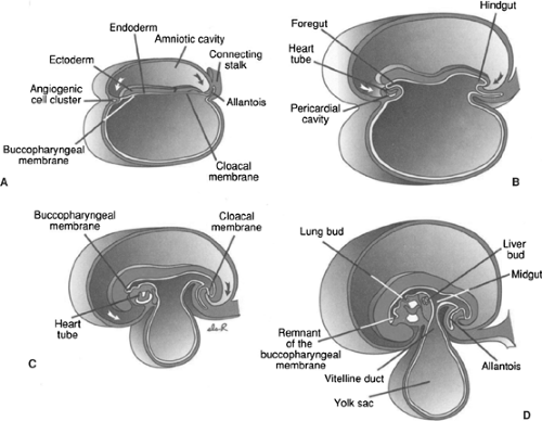
As a result of cephalocaudal and lateral folding of the embryo, a portion of the yolk sac cavity lined by the endoderm becomes incorporated into the embryo leading to the formation of the primitive gut. Two other portions of the cavity lined by endoderm, the yolk-sac and the allantois, remain outside the embryo (Fig. 128-1).
The primitive gut, extending from the cephalic to caudal parts of the embryo, forms a blind-ended tube comprising the foregut and hindgut. The middle portion or the midgut remains temporarily connected to the yolk sac by means of the vitelline duct, or yolk stalk (Fig. 128-1D). Further development of the primitive gut leads to the formation of the digestive tract. Endoderm forms the lining of the digestive tract and gives rise to the parenchyma or the glands such as the liver and pancreas, whereas muscle, connective tissue, and the peritoneal components of the wall of the gut are derived from splanchnic mesoderm.
The formation of the primitive gut and its derivatives into the digestive tract is usually defined into four distinct sections: (a) pharyngeal gut or pharynx, which extends from the buccopharyngeal membrane to the tracheobronchial diverticulum (Fig. 128-1D). This development is critical in the development of head and neck region. (b) The foregut is caudal to the pharyngeal gut and extends as far as the liver outgrowth. (c) The midgut begins caudal to the liver bud and extends to the junction of the right two-thirds and left third of the transverse colon in the adult. (d) The hindgut extends from the left third of the transverse colon to the cloacal membrane (Fig. 128-1).
The digestive system is initially formed by the lateral folding of the endodermal germ layer into a tube. From the simple tubular gut, development of the digestive system can be viewed at several levels, ranging from the elongation, herniation past the body wall, rotation, and folding for the effective packing into the body cavity. The digestive tube itself goes through the series of inductions and tissue interactions that provide the basis for development of the digestive glands, culminating in the biochemical maturation of the secretory and absorptive epithelia associated with the digestive tract. While these processes are occurring, the primordial digestive glands and respiratory structures are growing complex branching patterns as a result of continuous epithelial–mesenchymal interactions. In the developing digestive tube, regional mesenchymal influences determine the characteristics and functions of the epithelial lining specific to that region of the gut.
The structure and functions of the alimentary canal/digestive system range from those of esophagus to those of the rectum; however, these specialized regions have some of the basic structural organization that is similar throughout the length of the alimentary canal. The wall of the alimentary canal is formed of four distinct layers; from the lumen outward, these are as follows:
Mucosa: Consisting of an epithelial lining and an underlying connective tissue of the lamina propria and a layer of smooth muscle called the muscularis mucosa.
Submucosa: A layer of dense, irregular connective tissue.
Muscularis externa: A layer comprising both striated and smooth muscle.
Serosa or adventitia: A serous membrane consisting of a simple squamous epithelium, the mesothelium, and a small mound of underlying connective tissue. Where the wall is directly attached to adjoining structures, the outer layer is the adventitia, which is composed of connective tissue.
Development of the Esophagus
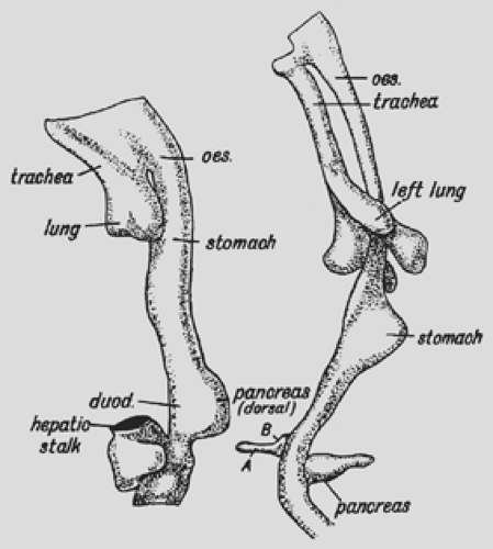
As early as the fourth week of development, the esophagus of the human embryo is merely a sphincter or constricted part of the primitive foregut, situated between the pharynx and stomach, as observed by Keith8 (Fig. 128-2).
During the sixth and seventh weeks of gestation, the esophagus undergoes rapid elongation as cephalic development separates the head and neck from the thorax. The elongation is facilitated by development of the lungs and pleural cavities, which push the stomach dorsally and inferiorly (Fig. 128-2). Keith suggests that the esophagus is of dual origin, with an upper retrotracheal part originating from the pharyngeal portion and an intratracheal part originating from the pregastric segment of the foregut. During the sixth week of development, the esophagus is only 2 mm long, but at birth it extends to 100 mm. Its superior limit is marked by the inferior cricopharyngeal portion of the inferior pharyngeal constrictor. Fyke and Code3 note that the
cricopharyngeal part of the inferior pharyngeal constrictor relaxes suddenly during swallowing and simultaneously lengthens the vocal folds of the larynx. The lower limit of the esophagus is marked by its entrance to the stomach, in a region that constitutes a barrier to reflux of gastric contents, but it is not marked by an anatomically recognizable sphincter. Basmajian1 has indicated that, at lower thoracic levels, the esophagus is supported away from the aorta, azygos vein, and body of the vertebrae, which permits advancement of the esophagus away from the vertebral column. As the lungs approach each other in an erect subject during inspiration, a retroesophageal window may be noted on radiographs (Fig. 128-2).
cricopharyngeal part of the inferior pharyngeal constrictor relaxes suddenly during swallowing and simultaneously lengthens the vocal folds of the larynx. The lower limit of the esophagus is marked by its entrance to the stomach, in a region that constitutes a barrier to reflux of gastric contents, but it is not marked by an anatomically recognizable sphincter. Basmajian1 has indicated that, at lower thoracic levels, the esophagus is supported away from the aorta, azygos vein, and body of the vertebrae, which permits advancement of the esophagus away from the vertebral column. As the lungs approach each other in an erect subject during inspiration, a retroesophageal window may be noted on radiographs (Fig. 128-2).
Keibel and Mall7 reported that as it reaches a length between 8.4 and 16 mm (at the fifth to seventh weeks), the esophagus is crescentic, with the concavity of the crescent directed toward the trachea (Fig. 128-2). Its upper part is round, but as it descends, it appears transversely elliptical. Near the level of tracheal bifurcation, it becomes round again; finally, it assumes an elliptical shape in the dorsoventral direction. The esophagus remains patent during this early period save for a nonspecific reticular coagulum.
Tissue Differentiation and Molecular Regulation During Development
Differentiation of various regions of the gut and its derivatives is dependent upon a reciprocal interaction between the endoderm (epithelium) of the gut tube and surrounding splanchnic mesoderm. Mesoderm dictates the type of structure that will form; for example, lungs in the thoracic region and descending colon from the hindgut region through a HOX code similar to the one that establishes the anterior (cranial) posterior (caudal) body axis. Introduction of this HOX code is a result of sonic hedgehog (SSH) expressed throughout the gut endoderm. Thus, in the region of the mid- and hindgut, expression of SSH in gut endoderm establishes a nested expression of the HOX code in the mesoderm. Once the mesoderm is specified by this code, it instructs the endoderm to form the various components of the mid- and hindgut regions, including the small intestine, cecum, colon, and cloaca (see Fig. 128-1). Similar interactions are responsible for partitioning the foregut.
Endoderm
The early work of Johnson6 and of Keibel and Mall7 was reviewed and supplemented by Johns.5 He reported that the esophagus of 3- to 6-mm embryos is lined by stratified columnar epithelium that is about three cells deep. Between 23 and 24 mm, the endoderm is two cells deep, followed by proliferation into many layers, which never occlude the esophagus but may narrow it at the level of tracheal bifurcation. Ciliated epithelium appears in the 40-mm embryo when ciliated cells form the superficial layers of the stratified columnar epithelium that lines the esophagus. This condition is characteristic of almost the entire esophageal lining up to the 130-mm stage. At this time, the stratified columnar condition is replaced by stratified squamous epithelium. Patches of ciliated epithelium may still be present in the upper part of the esophagus at birth. In the 62-mm embryo, patches of simple columnar epithelium appear in the upper and lower ends of the esophagus. Some columnar cells increase in height. Later, columnar cells are replaced by stratified ciliated columnar epithelium.
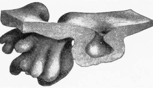
 Figure 128-3. Model of a superficial gland from the cardiac end of the esophagus of the human embryo at 240 mm (×120). The squamous epithelium of the slightly indented surface characterizes the morphology of this type of gland. (From Keibel F, Mall F. Manual of Human Embryology. Philadephia: Lippincott, 1912:364. With permission.)
Stay updated, free articles. Join our Telegram channel
Full access? Get Clinical Tree
 Get Clinical Tree app for offline access

|

